A Blade Got Detected Within the Head of a Dog
Received: 24-Oct-2022 / Manuscript No. jvmh-22-81066 / Editor assigned: 27-Oct-2022 / PreQC No. jvmh-22-81066 / Reviewed: 10-Nov-2022 / QC No. jvmh-22-81066 / Revised: 14-Nov-2022 / Manuscript No. jvmh-22-81066 / Published Date: 21-Nov-2022 QI No. / jvmh-22-81066
Abstract
An intracranial foreign body necessitated surgery for an 8-year-old Maltese dog. The dog had three generalised tonic-clonic seizures after being struck by an automobile. Using computed CT, an intracranial metallic item with a vertical orientation was discovered. The temporomandibular joint was in close proximity to the ventral end, which protruded by 2 mm. additionally; mild bilateral ventricular dilatation was noted. The foreign body was first attempted to be removed from the brain during surgery through the oral canal, but due to extensive covering by the surrounding tissues, a rostrotentorial craniotomy was ultimately decided. A sewing needle was discovered to be the alien object. The intracranial portion of the needle had been fully removed, according to postoperative computed tomography, leaving just the extracranial portion of the needle, which had separated from the remainder of the needle. Following surgery,antiepileptic medications were continued, and the dog is today symptom-free.
Background: In dogs and cats, a brain abscess or inflammation from a foreign body is a rare ailment. Skin, ear canals, conjunctiva, the nasal cavity, and oral cavity are all possible entry points for objects inside the skull. It is not unusual for clinical symptoms to appear a few weeks to months after the foreign object entered the patient’s body. The most frequent foreign bodies in human medicine are different surgical instruments, bullets, wood, nails, knives, and even sewing needles.The majority of people exhibit symptoms including headaches and seizures. Only seldom do conditions like cognitive loss, fever, brain abscesses, and meningitis occur, and roughly one-third of them are asymptomatic. 5 As a murderous effort, transcranial sewing needle penetration through the fontanelle occasionally happens (often related to cultural background). Up till 2010, 40 case reports regarding alleged sewing needle insertion via the fontanelle were published. 6 The majority of them had no clinical symptoms and infrequently had abnormal behaviour, headaches, or seizures. 5, 9 numerous case reports have been reported in which the child was pierced by the needle while still a newborn, but the clinical signs didn’t manifest until the child was an adult. Due to the potential risks of surgery, none
were necessary in the majority of these situations. 3, 6, 9, 10 A secondary growing brain abscess can sometimes elicit clinical indications in asymptomatic intracranial foreign entities after their penetration. In the veterinary literature,brain puncture by grass awns is a common occurrence. According to one investigation, an awn migration into the neurocranium via the orbital musculature led to a forebrain abscess. 1 All neurological symptoms (depressed mental status, spastic tetraparesis, and ataxia, with the pelvic limbs being more affected) were alleviated after the surgical
draining and extraction of the plant. A different kind of foreign body is a stick or piece of wood that can enter the brain and skull. In one case report, a stick was said to have entered a dog’s brain through the foramen ovale. In addition to the focused oral inflammation, neurological symptoms included abnormal mentation, no threat response in the left eye,and proprioceptive ataxia in all four limbs. An intracranial foreign body was detected using computed tomography (CT) imaging without the production of metallic artefacts. Based on this, a magnetic resonance imaging (MRI) examination was carried out, which revealed a long, hypointense linear structure encircled by a diffuse T2W hyperintensity on the right side of the brain. A haemorrhage was confirmed by the T2* sequence, and there were additional indications of cerebellar herniation, papilledema, and elevated intracranial pressure. After using an intraoral retrograde method to remove the stick, all clinical symptoms disappeared. The damaged part of the brain has a significant impact on the clinical characteristics. A 7-month-old dachshund with a brain abscess in the medulla oblongata caused ataxia, spastic tetraparesis, and a low mental status. 13 An MRI of the medulla revealed a circular mass that was T2W hyperintense and T1 hypointense, with contrast enhancement on T1W images of the lesion’s periphery. Euthanasia was decided because of the patient’s location and poor prognosis (diffuse glioma, abscess, etc.). The eventual conclusion following the postmortem examination was that a plant substance had caused the brain abscess. In addition to abscessation,meningoencephalitis and ventriculitis can be brought on by foreign materials, particularly grass awns. 14 These neurological abnormalities included extensor rigidity, altered mental state, tetraparesis or plegia, circling, and ataxia
in the three affected pups. Without using diagnostic imaging first, all of the dogs were put to sleep, and postmortem examination of one dog’s right occipital lobe and right lateral ventricle revealed serious symptoms of inflammation and a grass awn migration (another two dogs). 14 According to a case study, a juvenile Hungarian Vizsla experienced a seizure and gradually lost neurological function. 15 The right cerebral hemisphere displayed diffuse intra-axial T2W and fluid attenuated inversion recovery hyperintensity on the MRI, as well as a mass effect with a midline shift to the left. After the MRI, the dog’s spontaneous breathing did not return, therefore euthanasia was decided upon. Opening
the calvarium during postmortem inspection revealed a foreign body that resembled a plant in the caudal region of the right cerebral hemisphere (gyrus ectomarginalis medius). An study of the histopathology revealed perilesional pyogranulomatous meningoencephalitis. In the veterinary literature, a different kind of foreign body injury caused by porcupine quills to the central nervous system has been documented. 16 Injuries caused by quills can be frequent in areas where these animals are endemic, such as North America. One case report described a dog with sadness,halitosis, ptyalism, and oral pain brought on by an intracranial-intraaxial foreign body. 16 A CT scan indicated a foreign body in the dog’s brain, and the canine was put to sleep as a result of deteriorating clinical signs. A porcupine quill that had entered the skull through the foramen ovale was discovered during the postmortem examination.
Keywords
Blade; Head of a Dog; Surgery; Medications
Introduction
For needle removal surgery, an 8-year-old Maltese dog was referred to the Fuziovet Veterinary Referral Clinic and Hospital (Budapest, Hungary). The dog had a cluster seizure three days prior to the referral and had been involved in a small car accident. A nearby veterinarian took care of this, and oral phenobarbital was started as a long-term epilepsy treatment. The dog was recommended for a brain MRI test to [1-6] look at the potential underlying cause of the epilepsy. The neurological evaluation did not reveal any anomalies at the time of referral. Blood and urine tests, stomach and cardiac ultrasounds, and chest x-rays were routine preoperative examinations.
Materials &Methods
Investigations
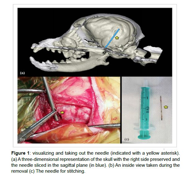
Metal was detected during scout imaging utilising a 1.5 T MRI (Siemens Magnetom Avanto, Siemens, Erlangen, Germany) scanner. The dog was moved from the MRI to a CT scanner (Siemens Somatom Sensation Cardiac CT, Siemens, Erlangen, Germany), which revealed an object with the appearance of a needle in the right side of the skull. This penetrated the bone and the temporal cortex and was situated rostral and medially to the temporomandibular joint at the lateralmost margin of the ala ossis basisphenoidalis (Figure 1). Additionally, somewhat dilated lateral ventricles were seen. The [7-10] squama temporalis, which is located in the middle ectosylvian gyrus, halted the needle’s tip dorsally. It’s interesting to note that the owner couldn’t recall any prior clinical evidence of needle swallowing. The dog never displayed any symptoms of mouth bleeding, decreased appetite, or oral pain.
Learning points/take-home messages
Even though it involves an additional step and costs more money, a doctor should consider a routine skull x-ray before sending a patient for a brain MRI if there is a suspicion of an intracranial foreign body.
This is because it has been shown that not all animals with intracranial metallic foreign bodies exhibit clinical symptoms. Due to the magnetic field’s action on the object in their case—especially a highfield magnetic resonance imaging—it could cause iatrogen trauma. Computed tomography testing is preferred if the x-ray shows that a metallic object is present. It may be sufficient in some circumstances to determine the disease’s origin and to make therapeutic options (e.g., conservative or surgical method). Additionally, it can be assessed how deeply the object is entrenched in the surrounding tissues or whether it is situated in a location where it is most likely to interact with the region of interest of a future magnetic resonance research. Low-field magnetic resonance imaging can be used if the computed tomography imaging results are insufficient (e.g., if further details need to be collected from the intracranial environment). Before doing the magnetic resonance scanning, a detailed cost-benefit analysis must be done because both the veterinarian and the owner should be aware of the potential risks. Preoperative planning for the surgical procedure should include both a decision tree outlining the potential surgical approaches and a threedimensional visualisation of the afflicted area to better comprehend the relationships (e.g., if the first route of intervention has to be changed intraoperatively, one should have an instant plan B to continue with). It should be highlighted that not all intracranial foreign objects can or should be removed based on case reports from human medicine. Thus, we emphasise the significance of multidisciplinary discussion and decision-making to select the most appropriate (conservative or surgical) approach for a particular instance.
Differential diagnosis
Seizures were the primary symptom in this case, which can be caused by structural brain damage from inflammation, tumours, congenital brain malformations, trauma, or secondary effects from systemic changes (reactive epilepsy); seizures can also have idiopathic causes. The lateral ventricles were found to be significantly enlarged on the CT scan, which can result in both epilepsy and brain damage. The metal foreign body inside the skull prevented the required MRI study from being done, thus we were unable to learn more about the brain or any potential changes brought on by head trauma. The probable (Figure 1) reasons of the symptoms were slight displacement of the foreign body due to the needle inside the brain, secondary brain damage, or trauma-related brain damage/hydrocephalus. The likelihood of an idiopathic origin was not completely ruled out, though.
Treatment
The needle detected by CT was removed from the dog at our facility. Three-dimensional (3D) reconstruction of the CT images was done prior to the operation. Using the 3D Slicer programme (https:// www.slicer.org), the osseous and metallic structures were separated out and exported as stereolithographic (STL) files. The models were improved using the Blender programme (https://www.blender. org). The dog was placed in sternal recumbency for the procedure, and a special tool was used to hold the head in place while opening the mouth. As a premedication, injections of fentanyl (5 mg/bwkg, Richter Gedeon), dormicum (0.05 mg/bwkg, EGIS), and ketamine (CP Ketamin 10% injectable AUV, Medicus Partner) were given. Propofol (1% MCT/LCT, 5.5 mg/bwkg, Fresenius-Kabi) was used to induce anaesthesia, and isoflurane and oxygen gas (1000 mg/g, 1.5 v/v%, Laboratorios Karizoo) were used to sustain it. An infusion pump was [10] used to continue the fentanyl-ketamine infusion (1 ml fentanyl + 0.06 ml ketamine/100 ml of infusion at an infusion rate of 100 ml/10 bwkg/h; fentanyl: 5 g/bwkg, Richter Gedeon; ketamine: CP ketamine 10% injection AUV, Medicus Partner) during the procedure.
Results &Discussion
In two dogs and a cat, the successful removal of a sewing needle from the brain has been documented. A domestic shorthair cat aged one had a needle inserted through the foramen magnum (from the atlas direction) to the brainstem. 2 The cat displayed a progression of clinical symptoms, starting with gingivitis and nasal discharge, then exhibiting behavioural issues, then experiencing issues with the mental state and conscious proprioception, along with a right-sided hemiparesis. All clinical symptoms were resolved once the needle was successfully removed from the caudal direction. A 11-month-old Cavalier King Charles spaniel had a sewing needle inserted through the foramen lacerum in one of the published case reports of dogs. 17 Hypersalivation, chewing, and a little increase in body temperature were the main clinical indicators. There was no discernible neurological deficit.
Conclusion
A metal-like object in the brain was discovered by radiographs and a CT scan, and it was extracted transorally under fluoroscopy supervision. Following the intervention, all clinical indicators vanished. A 1-year-old Maltese dog presented with hemorrhagic vomiting in the second reported occurrence, and 30 minutes after being referred, neurological indications appeared (hypermetric ataxia, proprioceptive deficit and progressive obtundation, status epilepticus).
Author Contributions
László Lehner and Kálmán Czeibert completed the data collection, data analysis, manuscript preparation, and article writing.
Acknowledgement
The authors appreciate VetScan for providing the preoperative CT pictures.
Conflict of Interest
There are no conflicts of interest, according to the authors.
Ethics Statement
This study did not need to be submitted to the local ethics and welfare council since all diagnostic studies and begun therapies were a regular component of clinical procedures.
References
- Cloquell A,Mateo I (2019)Surgical management of a brain abscess due to plant foreign body in a dog.Open Vet J 9:216–21.
- Cottam EJ,Gannon K (2015)Migration of a sewing needle foreign body into the brainstem of a cat.JFMS Open Rep1:1-10.
- Hao D,Yang Z,Li FA (2017) 61 year old man with intracranial sewing needle.J Neurol Neurophysiol8:1-10.
- Fischer BR,Yasin Y,Holling M,Hesselmann V (2012) Good clinical practice in dubious head trauma - the problem of retained intracranial foreign bodies.Int J Gen Med 5:899–902.
- Maghsoudi M,Shahbazzadegan B,Pezeshki A (2016)Asymptomatic intracranial foreign body: an incidental finding on radiography.Trauma Mon21:22206.
- Pelin Z,Kaner T (2012)Intracranial metallic foreign bodies in a man with a headache.Neurol Int.4:18.
- Sturiale CL,Massimi L,Mangiola A,Pompucci A,Roselli R,et al. (2010)Sewing needles in the brain: infanticide attempts or accidental insertion?Neurosurgery67:E1170–9.
- Abbassioun K,Ameli NO,Morshed AA (1979)Intracranial sewing needles: review of 13 cases.J Neurol Neurosurg Psychiatry.42:1046–9.
- Bozkurt H,Arac DA (2019) Late onset adult seizure due to intracerebral needle: case-based update.Childs Nerv Syst35:593–600.
- Sucu HK,Gelal F (2006)Intracranial metallic foreign body presenting with a unique route of introduction into the brain.Neurol India 54:224–5.
Indexed at, Crossref, Google Scholar
Indexed at, Crossref, Google Scholar
Indexed at, Crossref, Google Scholar
Indexed at, Crossref, Google Scholar
Indexed at, Crossref, Google Scholar
Indexed at, Crossref, Google Scholar
Citation: Wakgari MNS (2022) A Blade Got Detected Within the Head of a Dog. J Vet Med Health 6: 161.
Copyright: © 2022 Wakgari MNS. This is an open-access article distributed under the terms of the Creative Commons Attribution License, which permits unrestricted use, distribution, and reproduction in any medium, provided the original author and source are credited.
Select your language of interest to view the total content in your interested language
Share This Article
Recommended Journals
Open Access Journals
Article Usage
- Total views: 1476
- [From(publication date): 0-2022 - Nov 04, 2025]
- Breakdown by view type
- HTML page views: 1062
- PDF downloads: 414

