Vallecular Cyst: Airway Challenge and Options of Management
Received: 12-Jan-2014 / Accepted Date: 18-Feb-2014 / Published Date: 26-Feb-2014 DOI: 10.4172/2161-119X.1000158
Abstract
Vallecular cyst is a benign cyst with unknown aetiolgy. It is a rare presentation in adult and may obscure view for airway intubation. Prompt anticipation of difficult airway is important to reduce morbidity and mortality. We describe a case of vallecular cyst with surgical option for airway access and discuss the surgical management option based on current literature review.
Keywords: Asymptomatic vallecular cyst, Airway challenge
257331Introduction
Vallecular cyst is a benign cyst with unknown aetiolgy. It is a rare presentation in adult and may obscure view for airway intubation. Prompt anticipation of difficult airway is important to reduce morbidity and mortality. We describe a case of vallecular cyst with surgical option for airway access and discuss the surgical management option based on current literature review.
Case Report
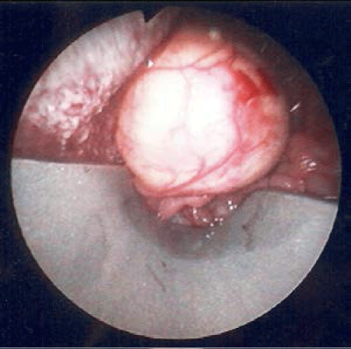
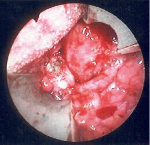
A 36years Malay gentleman presented to the ENT clinic with hoarseness of voice for the past one year. His symptoms were not associated with any difficulty in swallowing or breathing. He had history of tuberculosis treated a year earlier. On nasoendoscopy, there was a mass at the right vallecular region measuring 2 cm×3 cm. The vocal cord was normal. No other abnormality was identified. Routine blood and radiographic lateral neck x-ray did not reveal any abnormalities. MRI and CT SCAN of the neck demonstrated a lesion arising adjacent to the epiglottis seen at the region of hypopharynyx and vallecular measuring 2.7×2.6 cm. He consented for an excision biopsy of the cyst. On the operation table, endotracheal intubation via laryngoscope failed and emergency tracheostomy was performed to secure airway in view of the large vallecular cyst (Figure 3). Post operation, the histopathology report came back as a benign cyst (Figure 4).
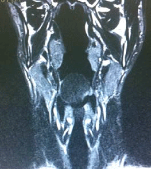
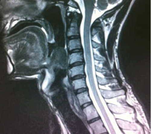
Axial view demonstrates lesion seen in the region of the hypopharynx and vallecular measuring 2.7 x 2.6 x 3.2 cm. It extends from the anterior aspect of epiglottis/base of tongue into the airway. Vocal cords appear normal (Figure 1). Sagital view shows the lesion arising adjacent to the epiglottis.This lesion also indents the posterior/ prevertebral wall causing narrowing of the airway. No obvious obliteration of the pyriform fossa. The hypo-pharynx appear normal (Figure 2).
Discussion
Vallecular cyst is a rare benign lesion which commonly arises from the lingual surface of the epiglottic region. It is known as epiglottic mucous retention, or base of the tongue cyst, and is classified as a ductal cyst that results from obstruction and retention of mucus in collecting ducts of submucosal glands containing clear and non-infected fluid [1]. This cyst is uncommon and its exact cause is unknown [1]. Several theories explained its pathogenesis. However two major hypotheses are that the cyst is a consequence of ductal obstruction or embryological malformation [2]. The commonest site of vallecula cyst is over the lingual surface of the epiglottis. Thus the increased size of the vallecular cyst may distort the epiglottis and fill the vallecular region as well as obstruct view of the airway [3]. This may lead to blockage of the laryngeal inlet and risk of respiratory airway distress [2,4].
There were a few methods on management of vallecular cyst. The conventional modalities include marsupialisation, or excision, where they were done either with CO2 laser or electrocautery using direct vision with or without microlaryngoscope with a camera assembly or via snaring of the cyst using a set of tonsillectomy instruments [5-9]. Depending on the size of the vallecular cyst difficult airway is a possibility in these patients. There were a few methods in current literature reviews for intubation options in patient presenting with vallecular cyst. The commonest is normal rapid sequence induction with cricoids pressure followed by oral intubation with the help of styletted endotracheal tube [6,8]. This is feasible if the size is small and no obstruction of view of the vocal cord. Another option is via transnasal fibreoptic intubation or flexible fibre optic nasolaryngoscopy. This can be used if the normal common endotracheal intubation fails [8]. The trachea can also be intubated via a rigid laryngoscope by the otolaryngologist, which can be used to displace the cyst to view the vocal cord as the rigid laryngoscope is longer than the anaesthetic laryngoscope [3]. The last option is usually tracheostomy performed by the ENT surgeon if all other methods fail [10]. In this case, intubation was unsuccessful via endotracheal intubation due to large vallecular cyst obstructing the view of the intubation. Thus an emergency tracheostomy was performed by the ENT surgeon before surgical procedure of removing the cyst. Post operative period was uneventful and the patient was discharged on the 3rd post operative day after weaning down fromthe tracheostomy. On follow up one week later, via endoscopic review, the wound was found to have healed completely and no recurrence noted.
This case reinforces the need to avoid repeated intubation attempts as this may increase complications as well as the need for an ENT surgeon to prepare for tracheostomy should endotracheal intubation fails via normal methods. Repeated attempts at intubation following difficulty to visualize the vocal cord may predispose patient to airway distress or cyst rupture or even aspiration.In opting for tracheostomy to secure the airway in this case, complications of failed intubation could be avoided.
Conclusion
In preparing a patient with vallecular cyst for surgery, meticulous airway assessment and proper planning are mandatory as any difficult airway scenarios.Pre-operative assessment should be meticulously made in determining the options of intubation. Tracheostomy should be planned in advance if the vallecular cyst obstructs visualization of the airway.
References
- Parelkar SV, Patel JL, Sanghvi BV, Joshi PB, Sahoo SK, et al. (2012) An Unusual Presentation of Vallecular Cyst with near Fatal Respiratory Distress and Management Using Conventional Laparoscopic Instruments. J Surg Tech Case Rep 4: 118-120.
- Sanjeev Mohanty, Gopinath Maraignanam (2013) Endoscopic assisted therapeutic marsupialisation of vallecular cyst in a seven year old boy- a rarity in modern clinical practice.Journal of Evolution of Medical and Dental Sciences 2: 4320-4324.
- C M Walshe, N Jonas, DRohan (2009) Vallecular cyst causing a difficult intubation. Oxford University Press on behalf of The Board of Directors of the British Journal of Anaesthesia 102: 565.
- Berger G, Averbuch E, Zilka K, Berger R, Ophir D (2008) Adult vallecular cyst: thirteen-year experience. Otolaryngol Head Neck Surg 138: 321-327.
- Rivo J, Matot I (2001) Asymptomatic vallecular cyst: airway management considerations. J ClinAnesth 13: 383-386.
- Sameer M, Jahagirdar, P Karthikeyan, Ravishankar M (2013) Acute Airway Obstruction, an unusual presentation of vallecular cyst. Indian J Anaesth 55: 524-527.
- SathishBhandary (2003) Case report: Innovative Surgical Technique in the Management of Vallecular cyst JHAS Mangalore, South India 2:2.
- Kothandan H, Ho VK, Chan YM, Wong T (2013) Difficult intubation in a patient with vallecular cyst. Singapore Med J 54: e62-65.
- SinghalSurinder K, Verma Hitesh, Dass Arjun, PuniaRajpal (2012) Vallecular cyst in Adult Population: Ten Year Experience. Nepalese Journal of ENT Head and Neck Surgery 2.
- Al-Qudah M, Shetty S, Alomari M, Alqdah M (2010) Acute adult supraglottitis: current management and treatment. South Med J 103: 800-804.
Citation: Nee TS, Saim A (2014) Vallecular Cyst: Airway Challenge and Options of Management. Otolaryngology 4:158. DOI: 10.4172/2161-119X.1000158
Copyright: © 2014 Nee TS. This is an open-access article distributed under the terms of the Creative Commons Attribution License, which permits unrestricted use, distribution, and reproduction in any medium, provided the original author and source are credited.
Share This Article
Recommended Journals
Open Access Journals
Article Tools
Article Usage
- Total views: 26237
- [From(publication date): 5-2014 - Apr 02, 2025]
- Breakdown by view type
- HTML page views: 21527
- PDF downloads: 4710


