Pott's Puffy Tumor Complicating Frontal Sinus Osteoma - A Case for Combined Approach Surgery
Received: 12-Nov-2013 / Accepted Date: 30-Dec-2013 / Published Date: 07-Jan-2014 DOI: 10.4172/2161-119X.1000152
255518Introduction
Paranasal sinus osteomas are slow-growing osteogenic tumours named after the sinus invaded, with the frontal sinus most commonly involved [1]. Typically, frontal sinus osteomas are asymptomatic and coincidentally found in up to 3% of imaging [2]. Unless symptomatic or complicated, these do not require surgery. Sinus osteomata can result in obstructive disease with its associated orbital and intracranial sequelae [3,4].
Some authors suggest surgical intervention if over 50% of the frontal sinus is occupied by an osteoma [5]. The ideal surgical approach aims to achieve restoration of the drainage pathways, with endonasal, external and combined approaches described. External approaches are associated with risk of sinocutaneous fistulation and sub-optimal management of underlying sinusitis. This has led to interest in development of endonasal approaches. Where access to the anterior table is limited with the endoscope alone, a combined approach is necessary to fully eradicate the disease.
Pott’s Puffy Tumor (PPT) describes a subperiosteal abscess overlying frontal bone osteomyelitis which presents as a painful, fluctuant forehead tumor. It is a rare complication, its most common cause being frontal sinusitis. One-third of PPT is associated with lifethreatening intracranial complications [6]. Causative organisms are those frequently implicated in chronic rhinosinusitis: streptococci, staphylococci and anaerobes [7]. Optimum management involves draining the collection, restoring sinus ventilation, debriding the osteomyelitic bone and granulation tissue and protracted antibiotic therapy guided by microbiologist advice [8].
We present the first case of Pott’s puffy tumor caused by a frontal sinus osteoma and describe the use of a combination of external and endoscopic approaches in managing this condition. The challenge in both this case, and in successful management of frontal sinus osteoma and PPT, is eradication of the disease, restoration of sinus ventilation and prevention of recurrence – a case for combined approach surgery.
Case Report
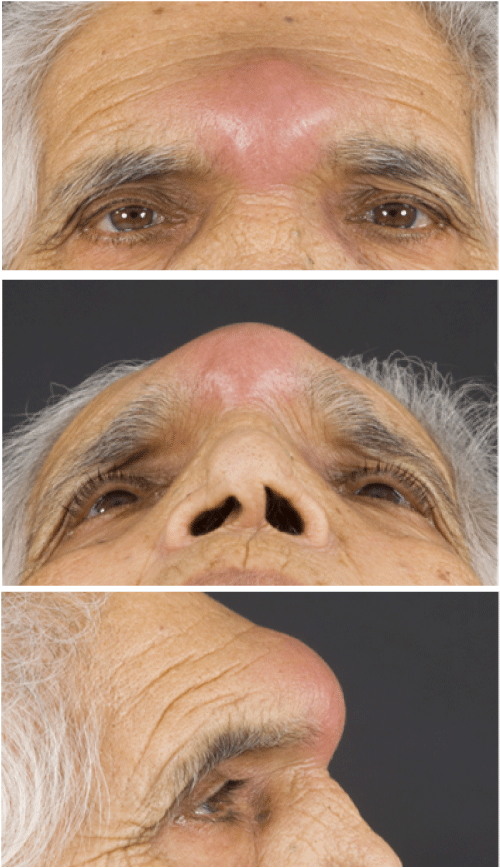
An 85 year old female presented to our tertiary centre with a sixmonth history of progressive forehead swelling refractory to multiple courses of oral flucloxacillin, metronidazole and co-amoxiclav. Symptoms of vertex headache, facial pain, epiphora and pyrexia were described. Examination confirmed a tense, fluctuant forehead swelling (Figure 1). Fibreoptic nasendoscopy revealed a grade I polyp in the left middle meatus with normal appearance of the nasal mucosa otherwise. There were no signs of meningism, cranial nerve palsy or neuroophthalmic deficit.
Initial investigations of full blood count, bone profile and urea & electrolytes were normal. C-reactive protein was elevated at 49 g/dL and this gradually normalized after surgery and targeted intravenous antibiotic therapy. Empirical antibiotic therapy guided by microbiology advice was initiated. Tazocin (piperacillin and tazobactam) was selected for its extended spectrum of activity against both Gram negative and Gram positive pathogens, and anaerobes including Pseudomonas aeruginosa; metronidazole was chosen for its complementary additional anaerobic cover and action against Protazoa. Antibiotic choice was revised at the time of surgery to meropenem, for its similar coverage spectrum and ability to cross the blood-brain barrier.
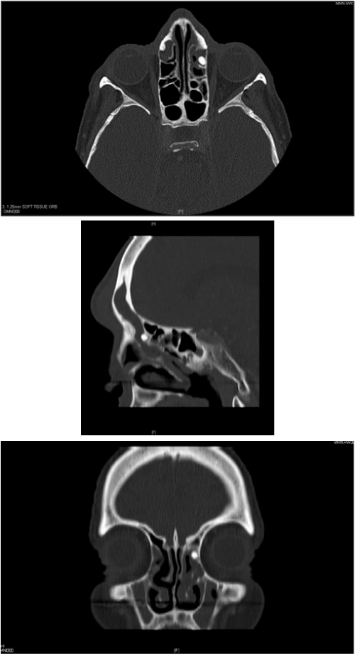
HRCT confirmed PPT with an anterior table defect of the left frontal sinus, and an osteoma narrowing the ipsilateral frontal recess. The posterior table remained intact (Figure 2).
The abscess was drained via a combined approach: the osteoma obstructing the frontal recess was debrided endoscopically and an external approach via a small Lynch Howarth incision was performed. Tissue biopsy and pus were taken for culture, sensitivity, histopathology, mycology and Acid-Fast Bacteria (AFB) staining. A drain placed in the frontal sinus was left in situ for the purpose of daily irrigation. There were no surgical complications.
Histology revealed necrotizing vasculitis of the frontal sinus tissue in the absence of autoimmune serology, and bony fragments with no features of malignancy were demonstrated in the osteoma specimen. Strepcoccus sanguinis was cultured and symptoms resolved after one week of intravenous meropenem. Oral co-amoxiclav was continued for four weeks on microbiologist advice.
The histological presence of a vasculitic process of the frontal sinus prompted liaison with Rheumatologists, and serological investigations and urinalysis for red cell casts were performed. Results of these investigations suggested the presence of vasculitic glomerulonephritis. The patient was unable to attend for follow-up due to relocation abroad.
Discussion
In this unusual case the causative factor of Pott’s puffy tumor was a frontal sinus osteoma which required excision to restore sinus ventilation and drainage pathways, and to eradicate active disease. Failure of removal of the osteoma would result in persistent obstruction of the frontal sinus and disease would likely recrudesce.
A combined endonasal and external approach was necessary to achieve these aims. The external Lynch Howarth incision provided direct access for pus drainage, debridement and tissue biopsy, and an externally-placed drain allowed for regular irrigation of the frontal sinus. Osteoplastic flap was not required as the low-grade osteoma was accessible endoscopically.
Targeted antibiotic therapy, identification of a vasculitic role in the aetiology and confirmation of an osteoma as the primary cause in this patient were guided by both tissue and pus samples obtained for microbiology, histology and gram stain.
Pott’s puffy tumour
The mainstay of treatment for PPT is drainage; debridement and a protracted course of broad spectrum antibiotic treatment [8]. In the post-antibiotic era supra-additive causative pathologies may be involved in the development of PPT. It is therefore prudent to provide both tissue and pus samples for laboratory analysis.
In a review of 22 cases Verbon et al. found a preponderance of PPT in adolescents, and of the few adults affected most had either negative or atypical pus culture results [7]. Other reported unusual causes include Pasteurella multocida which is commonly associated with animal bites or asymptomatic colonisation in chronic respiratory disease, mastoiditis, mucormycosis, association with maxillary rather than frontal sinusitis and association with frontal sinusitis secondary to dental sepsis [8-14].
Osteoma
Endonasal (Draf IIA, IIB, III/modified Lothrop procedure) external and combined approaches have been described in the removal of symptomatic frontal sinus osteomas [14].
Chiu et al. [15] proposed a grading system of the frontal sinus osteomas to guide surgical management (Table 1), with recommendation that low grade osteomas (I + II) are endoscopically managed and high grade lesions (III + IV) are removed via an external approach with osteoplastic flap.
| Grade | Characteristics |
|---|---|
| I | Origin of the osteoma posterior and inferior in the frontal recess; localization of osteoma medial to a virtual sagittal plane passing through the orbital lamina; Anteroposterior (AP) diameter of the tumour is < 75% of the AP diameter of the frontal sinus |
| II | As grade I; AP diameter of the tumour> 75% of the AP diameter of the frontal sinus. |
| III | Origin of the osteoma anterior and/or superior in the frontal sinus and/or osteoma extending lateral to a virtual sagittal plane passing through the orbital lamina. |
| IV | Osteoma fills the entire frontal sinus. |
Table 1: Grading of osteomas as devised by Chiu et al. [15].
Bi-coronal and Lynch Howarth incisions with osteoplastic flap are advantageous in direct visualisation of the frontal sinus in advanced disease. In a case series of seventeen patients Al-Qudah and Graham found osteoplastic flap use was indicated in lateral disease [16]. However there is risk of morbidity, scarring and sinocutaneous fistulation with external approach. There is also risk of inadvertent injury to intracranial or ophthalmic structures, for example where there is large orbital involvement, invasion of the anterior or posterior table, or extension intracranially or far laterally beyond the orbital meridian.
In a retrospective review of patients who underwent endonasal surgery for low grade osteomas of the frontoethmoidal junction and frontal sinus, Pagella et al. [17] report successful access medial to the sagittal plane of the lamina papyracea and to the frontal recess. This was aided by a long handpiece, curved endonasal drills and intraoperative image-guidance. This is consistent with the case presented, though in the experience of the authors visualisation may be challenging in the presence of inflamed mucosa, thus limiting access to the anterior table.
As demonstrated by Castelnuovo et al. [1] in a comparison with the open approach, endonasal surgery confers shorter post-operative stay and increased patient comfort. The development of technology has facilitated endoscopic removal of high grade osteomas. This is presented in series by Sieberling et al. [18] and Ledderose et al. [19] who demonstrated successful removal of large osteomas using Draf III / modified Lothrop procedure. A proportion of cases in these series required a combined approach or remnants of residual disease (Ledderose: two of fifteen; Sieberling: nine of fourteen).
When considering endoscopic removal of a frontal sinus osteoma the size, position of the lesion, obstructive features, its relationship to neural structures and the experience of the surgical team and available resources should be taken into account [20].
Conclusions
Frontal sinus osteoma may result in PPT. The clinician ought to consider this both when treating this under-reported complication of frontal sinusitis, and when managing the patient with frontal sinus osteoma. Thorough and timely treatment of PPT is essential to decrease the risk of intracranial sepsis and targeted therapy should be guided by culture, histological analysis and gram stain of pus and tissue samples.
Endoscopic and external approaches to the frontal sinus are not mutually exclusive and can complement one another. Such as the case presented, a combined approach could be necessary to most effectively treat the disease process and these cases may warrant referral to a tertiary care facility. Experience of combined approaches and endoscopic removal of high grade osteomas should be shared to accrue evidence of management in these challenging cases.
References
- Castelnuovo P, Valentini V, Giovannetti F, Bignami M, Cassoni A, et al. (2008) Osteomas of the maxillofacial district: endoscopic surgery versus open surgery. J Craniofac Surg 19: 1446-1452.
- Earwaker J (1993) Paranasal sinus osteomas: a review of 46 cases. Skeletal Radiol 22: 417-423.
- GuedesBde V, da Rocha AJ, da Silva CJ, dos Santos AR, Lazarini PR (2011) A rare association of tension pneumocephalus and a large frontoethmoidalosteoma: imaging features and surgical treatment. J Craniofac Surg 22: 212-213.
- Sakamoto H, Tanaka T, Kato N, Arai T, Hasegawa Y, et al. (2011) Frontal sinus mucocele with intracranial extension associated with osteoma in the anterior cranial fossa. Neurol Med Chir (Tokyo) 51: 600-603.
- Smith ME, Calcaterra TC (1989) Frontal sinus osteoma. Ann Otol Rhinol Laryngol 98: 896-900.
- Singh B, Van Dellen J, Ramjettan S, Maharaj TJ (1995) Sinogenic intracranial complications. J Laryngol Otol 109: 945-950.
- Verbon A, Husni RN, Gordon SM, Lavertu P, Keys TF (1996) Pott's puffy tumor due to Haemophilusinfluenzae: case report and review. Clin Infect Dis 23: 1305-1307.
- Ketenci I, Unlü Y, Tucer B, Vural A (2011) The Pott's puffy tumor: a dangerous sign for intracranial complications. Eur Arch Otorhinolaryngol 268: 1755-1763.
- Skomro R, McClean KL (1998) Frontal osteomyelitis (Pott's puffy tumour) associated with Pasteurellamultocida-A case report and review of the literature. Can J Infect Dis 9: 115-121.
- Khan MA (2006) Pott's puffy tumor: a rare complication of mastoiditis. Pediatr Neurosurg 42: 125-128.
- Effat KG, Karam M, El-KabaniA (2005) Pott's puffy tumour caused by mucormycosis. J Laryngol Otol 119: 643-645.
- Maheshwar AA (2001) Pott’s puffy tumor: an unusual presentation and management. Journal of Laryngology and Otology 115: 497-499.
- Chandy B, Todd J, Stucker FJ, Nathan CO (2001) Pott's puffy tumor and epidural abscess arising from dental sepsis: a case report. Laryngoscope 111: 1732-1734.
- Draf W (2005) Endonasal frontal sinus drainage type I-III according to Draf. In Kountakis S, Senior B, Draf W, editors. The Frontal Sinus, 1st edition, New York. Springer p 219-230.
- Chiu AG, Schipor I, Cohen NA, Kennedy DW, Palmer JN (2005) Surgical decisions in the management of frontal sinus osteomas. Am J Rhinol 19: 191-197.
- Al-Qudah M, Graham SM (2012) Modified osteoplastic flap approach for frontal sinus disease. Ann Otol Rhinol Laryngol 121: 192-196.
- Pagella F, Pusateri A, Matti E, Emanuelli E (2012) Transnasal endoscopic approach to symptomatic sinonasalosteomas. Am J Rhinol Allergy 26: 335-339.
- Seiberling K, Floreani S, Robinson S, Wormald PJ (2009) Endoscopic management of frontal sinus osteomas revisited. Am J Rhinol Allergy 23: 331-336.
- Ledderose GJ, Betz CS, Stelter K, Leunig A (2011) Surgical management of osteomas of the frontal recess and sinus: extending the limits of the endoscopic approach. Eur Arch Otorhinolaryngol 268: 525-532.
- Lund VJ, Stammberger H, Nicolai P, Castelnuovo P, Beal T, et al. (2010) European position paper on endoscopic management of tumours of the nose, paranasal sinuses and skull base. Rhinol Suppl : 1-143.
Citation: Khan MM, Khwaja S, Bhatt YM, Karagama Y (2014) Pott’s Puffy Tumor Complicating Frontal Sinus Osteoma - A Case for Combined Approach Surgery. Otolaryngology 4:152. DOI: 10.4172/2161-119X.1000152
Copyright: © 2014 Khan MM, et al. This is an open-access article distributed under the terms of the Creative Commons Attribution License, which permits unrestricted use, distribution, and reproduction in any medium, provided the original author and source are credited.
Share This Article
Recommended Journals
Open Access Journals
Article Tools
Article Usage
- Total views: 18469
- [From(publication date): 2-2014 - Apr 03, 2025]
- Breakdown by view type
- HTML page views: 13897
- PDF downloads: 4572


