‘New Developed Femoral-S Carotid Catheter’ is Very Effective and Safe in Carotid Angiography: A Retrospective Study
Received: 10-Nov-2017 / Accepted Date: 28-Nov-2017 / Published Date: 05-Dec-2017 DOI: 10.4172/2167-7964.1000274
Abstract
Objective: There is no special catheter used for carotid angiography in the market. We studied the efficacy feasibility and safety of Femoral S carotid catheter (made by shaping existing catheters) in this study.
Methods: 1169 patients undergoing transfemoral or transradial carotid angiography between the years 2015 and 2017 were evaluated. The 156 patients found that are underwent carotid angiography with Femoral S catheter.
Results: The Baseline characteristics of the patients were record. The total time of the procedure with the new catheter was found 7.2 ± 1.3 min and the amount of opaque media used was 50.20 ± 15.60 ml. Selective imaging success was high 95%. Minor Complication (local hematoma, leg pain, dizziness, headache, hypotension, allergic reaction of the contrast media) development was found 3.8%. Permanent damage, morbidity and/or mortality were not seen in patients.
Discussion: The right judkins catheter is usually preferred in the carotid angiography performed with the femoral route. The newly developed catheter does not stick to the aortic wall because the tip is in the “S” shape. Therefore, the processing time is shorter and the imaging performance is better than multipurpose catheter.
Conclusion: Carotid angiography with the newly developed Femoral S carotid catheter seems easier and safer. Performing of Carotid angiography may help to reduce stroke frequency at the time of coronary angiography in patients with coronary artery disease.
Keywords: Carotid angiography; New carotid catheter; Stroke; Multipurpose catheter
Introduction
As is known, the association of coronary atherosclerosis with peripheral atherosclerosis is very common. This frequency is predictive of the likelihood of someone from stroke, kidney failure, and limb ischemia in the people with coronary artery disease (CAD). It may be beneficial for the patient to look at other possible atherosclerotic vessels at the moment of coronary angiography.
The Invasive carotid angiography is also needed for diagnosis and treatment of neck malformations, carotid tumours, vascular malformations and injuries as well as USG, MR angiography, CT angiography [1-6].
Catheter reshaping is an ancillary method used in catheter laboratories, to increase success in imaging modalities. The number of physicians who use this method to display vessels with origin abnormality is steadily increasing. In our laboratory, we also use catheter reshaping since long time for cases where the existing catheters are inadequate for the imaging of vessels with origin anomalies [7,8].
Method
Since 2009, we have performing coronary angiography followed by carotid angiography with heart diseases that are we suspect of carotid artery disease to the patients who signed informed consent form(those who had, suspicious carotid artery stenos in Carotid Doppler USG, those who had stenos in the USG, whose murmur was heard in the neck, and stroke in his history).
Carotid artery and coronary angiography were not performed on patients those who had, kidney failure, the same limb ischemia, no femoral pulses.
In addition, no carotid angiography was performed at risky persons (Those who had severe plaque in the carotid ostia, severe atherosclerotic calcified aortic cases, severe skeletal anomalies, major thoracic injuries, aortic surgery, aortic aneurysms with a diameter is larger than 5.0 cm).
156 patients underwent carotid angiography with a newly developed Femoral S catheter. The all patient data and the results were transferred to SPSS and analysed.
Angiography protocol
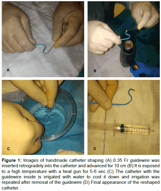
The femoral region was disinfected and local anaesthesia was performed with 10 cc, 0.05 prilocain. The femoral artery was cannulated with Terumo 6f sheet. The left coronary artery was visualized with a JL catheter, the right coronary artery was visualized with a JR catheter, and then the carotids were imaged with a Femoral S catheter in the patients. A 6F multipurpose catheter was used to generate each Femoral S catheter. The Reshaping was done in the lab at the time of operation (Figure 1).
Figure 1: Images of handmade catheter shaping (A) 0.35 Fr guidewire was inserted retrogradely into the catheter and advanced for 10 cm (B) It is exposed to a high temperature with a heat gun for 5-6 s (C) The catheter with the guidewire inside is irrigated with water to cool it down and irrigation was repeated after removal of the guidewire (D) Final appearance of the reshaped catheter.
Catheter shaping and carotid angiography
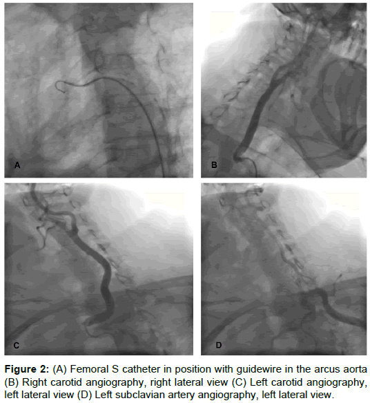
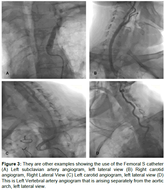
Distal tip of the 0.035 guide-wire we used in angiography was inserted through distal tip of the catheter, and advanced for 10 cm, and then with the aid of the guide-wire the required shaping was performed. The desired styling was done, afterwards the end of the catheter was held 10 cm away from the heat gun and exposed to hot air for 4-6 s, Immediately after, the Heated and shaped catheter tip was inserted into sterile isotonic water, Using a plastic injector, the catheter with the guide-wire inside was irrigated from its opposite end with sterile isotonic saline solution. The wire was removed and cooling with water was continued before the cooling was completed. As the catheter cooled, it was advanced to the arcus aorta with the help of a 0.35 guide-wire. The carotids were found and displayed with catheter rotation (Figures 1-3).
Figure 3: They are other examples showing the use of the Femoral S catheter (A) Left subclavian artery angiogram, left lateral view (B) Right carotid angiogram, Right Lateral View (C) Left carotid angiogram, left lateral view (D) This is Left Vertebral artery angiogram that is arising separately from the aortic arch, left lateral view.
Angiographic data of all patients, laboratory results, and registered epicrisis data were retrospectively reviewed. All the data were analysed after loading the SPSS.
Statistical analysis
Continuous variables are presented as mean ± standard deviation and nominal variables as numbers and percentages. A 2-sided P-value below 0.05 was considered significant.
Results
The Baseline characteristics are recorded. In the patients of femoral S catheter (n: 156), the procedure time was found mean 7.2 ± 2.3 min and the amount of opaque used 50 ± 15 ml. The incidence of minor complications was found (n=6) 3.8%. Major complications, morbidity and mortality were not observed in our patients (Tables 1 and 2).
| Femoral S (n: 156) |
|
|---|---|
| Female gender | 52 (33%) |
| Age | 67.9 ± 8.9 |
| Hypertension | 145 (92.9%) |
| Diabetes mellitus | 63 (42%) |
| Prior stroke or TIA | 65 (41.7%) |
| Prior PCI | 94 (60%) |
| Prior CABG | 37 (23.7%) |
| Hyperlipidemia | 121 (76%) |
| Renal failure | 0 |
Table 1: Baseline characteristics of patients.
| Femoral S (n: 156) |
|
|---|---|
| Total procedure time (min) | 7.2 ± 1.3 |
| Amount of contrast media used | 50.2 ± 15.6 |
| Selective Imaging | 152 (95%) |
| CAD with Carotid artery Disease | 124 (79%) |
| Major complication, CVA or Death | 0 |
| All minor complications | 6 (3.8%) |
Table 2: Results.
Selective imaging success was significantly high in the procedure (95%). Non-selective imaging is not as good as selective imaging, but it is enough to show carotid stenos. However, selective imaging success was found to be significantly high in the procedure with Femoral S catheter (Table 2).
Arcus aorta types and frequencies
Cardiologists who have performing carotid angiography should know the types of the arcus aorta, which is important for procedural safety and success. Some publications have different classifications for arcus aorta types. Most of these classifications are insufficient to help carotid angiography. The most appropriate classification for us is the classification of Huapaya JA and Ergun E.
The arcus aorta types we encounter in our cases and their frequencies are close to the classification of Ergun E. Type I is the most common type, known as the normal arcus aorta, occurring in 86% of cases. In this type of arcus aorta, the right carotid artery is separated from the right brachiocephalic artery, while the left carotid artery leaves the arcus aorta directly, after 1-2 cm from the brachiocephalic artery. Type 2 is found around 9%; Right and left common carotid artery exit from the brachiocephalic artery. In Type 3, the right and left common carotid arteries come out directly or separately from the arcus aorta in a ratio of 2%. Type 4 is including arteria lusoria, the right subclavian artery is removed from the aorta after the carotid arteries. This type of aorta is seen in 3% of the population [9-11].
Discussion
There is a strong correlation between coronary atherosclerosis and cerebral and carotid atherosclerosis. Carotid imaging at the moment of coronary angiography is an opportunity for the patient. CT angiography, MR angiography may show the degree of the narrowing of the carotid artery, but is not as satisfactory as invasive angiography on the about assessing the presence of thin fissures. They cannot correctly display the plaque character, whether it is sensitive plaque or not, the presence and severity of plaque cracks. For all these reasons, invasive angiography in carotid imaging is still the gold standard [2,12-15].
Most of the available angiography catheters have been designed for the femoral access. Carotid angiography can be performed with the Judkins Right catheter, one of these catheters. Nevertheless, the complication can be seen more with inexperienced hands and conventional techniques. This situation discourages physicians from making carotid angiography [16-18].
There are publications about the investigating reliability of transradial coronary and carotid angiography and their advantages over the femoral access. However, carotid imaging with the transradial approach is more difficult and requires more experience. Therefore, the first preferred route for carotid angiography is the femoral way. New catheter development is needed for physicians to making easier and safer carotid imaging with femoral way [19-26].
The Femoral S catheter that we have developed for this purpose seems to be more advantageous than the JR catheter. The catheter tip does not frict the aortic wall when rotating this catheter. Thanks to the “S” shape of our new catheter, it is possible to maneuver without 0.035 guide-wire and without encountering any resistance. Thanks to the advantages of our catheter, the procedure time is shortening and opaque consumption reducing. However, the tips of JR catheter can friction to the aortic wall if it is rotated within the aorta without guidewire. This condition can cause complications [1,15].
Study limitations
The goal in this study is to demonstrate the success of a hand-made catheter on a carotid angiography. A comparative study of femoral S catheter with the Simon, HN4-5, CK1 and man catheters would have been much more demonstrative. However, in our Web-based scan, we could not find any studies showing that these catheters were successful in carotid angiography. Therefore, these catheters are suitable catheters those can be used in the carotid imaging with the femoral route
Conclusion
Concomitant carotid angiography with coronary angiography may be an advantage for patients with coronary artery disease, because people with coronary artery disease are likely to having carotid artery disease.
Making of Carotid angiography is difficult with existing catheters for selectively viewing carotid artery. The newly developed catheter is more successful than multipurpose catheters in the carotid angiography performing. The procedure time is shorter with new catheter and the amount of opaque used is lesser. The quality of carotid images is better with the new catheter because more selective carotid angiography can be done with this catheter. Safe and more satisfyingly performing of carotid angiography will be more beneficial to the physician and the patient.
Disclosures
The author confirms that there are no known conflicts of interest associated with this publication and there has been no significant financial support for this work that could have influenced its outcome.
References
- Rigatelli G (2005) Screening angiography of supraaortic vessels performed by invasive cardiologists at the time of cardiac catheterization: Ä°ndications and results. Int J Cardiovasc Imaging 21: 179-183.
- Rigatelli G, Gemelli M, Zamboni A, Docali G, Rossi P, et al. (2003) Significance of selective carotid angiography during complete cardiac catheterization in patients candidates to combined aortic valve and carotid surgery. Minerva Cardioangiol 51: 305-309.
- Rigatelli G (2004) Diagnosis of carotid artery occlusive disease in patients scheduled for cardiac or vascular surgery: Ä°s this a place for invasive selective carotid angiography? Stroke 35: e89-e90.
- Korotkikh NG, Ol'shanskiÄ MS, Stepanov IV (2011) Multidisciplinary approach to diagnostics of extensive vascular head and neck malformations. Stomatologiia 91: 40-45.
- Korotkikh NG, Ol'shanskiÄ MS, Stepanov IV, Mashkova TA, NerovnyÄ AI, et al. (2013) The multidisciplinary approach to diagnostics and treatment of hypervascular masses in ENTstructures. Vestn Otorinolaringol 5: 44-47.
- Arslan F, Yılmaz S, Özer F, Andıç C, Canpolat T, et al. (2012) Surgical treatment of carotid body tumors. Kulak Burun Bogaz İhtisas Dergisi 23: 336-340.
- Erden I, Golcuk E, Bozyel S, Erden EC, Balaban Y, et al. (2017) Effectiveness of handmade “Jackyâ€Like Catheter†as a single multipurpose catheter in transradial coronary angiography: A randomized comparison with conventional twoâ€catheter strategy. J Ä°nterventional Cardiol 30: 24-32.
- Kim JY, Yoon SG, Doh JH, Choe HM, Kwon SU, et al. (2008) Two cases of successful primary percutaneous coronary intervention in patients with an anomalous right coronary artery arising from the left coronary cusp. Kor Circul J 38: 179-183.
- Huapaya JA, Chávez-Trujillo K, Trelles M, Dueñas Carbajal R, Espadin FR (2015) Anatomic variations of the branches of the aortic arch in a Peruvian population. Medwave 15: e6194.
- Ergun E, Şimşek B, Koşar PN, Yılmaz BK, Turgut AT (2013) Anatomical variations in branching pattern of arcus aorta: 64-slice CTA appearance. Surg Radiol Anat 35: 503-509.
- Cummings MS, Kuo BT, Ziada KM (2011) A rare anomaly of the aortic arch: aberrant right subclavian artery associated with common carotid trunk. J Invasive Cardiol 23: E241-E243.
- Seo WK, Yong HS, Koh SB, Suh SI, Kim JH, et al. (2008) Correlation of coronary artery atherosclerosis with atherosclerosis of the intracranial cerebral artery and the extracranial carotid artery. Eur Neurol 59: 292-298.
- Cha KS, Kim MH, Kim YD, Kim JS (2001) Combined right transradial coronary angiography and selective carotid angiography: safety and feasibility in unselected patients. Catheter Cardiovasc Ä°nterv 53: 380-385.
- Rafailidis V, Chryssogonidis I, Tegos T, Kouskouras K, Charitanti-Kouridou A (2017) Imaging of the ulcerated carotid atherosclerotic plaque: A review of the literature. Insight Ä°mag 1-13.
- Ledwoch J, Staubach S, Segerer M, Strohm H, Mudra H (2017) Incidence and risk factors of embolized particles in carotid artery stenting and association with clinical outcome. Int J Cardiol 227: 550-555.
- Berczi V, Randall M, Balamurugan R, Shaw D, Venables GS, et al. (2006) Safety of arch aortography for assessment of carotid arteries. Eur J Vasc Endovasc Surg 31: 3-7.
- Morgan JH, Johnson JH, Brown RB, Harvey RL, Rizzoni WE, et al. (2006) Initial experience with routine selective carotid arteriography by vascular surgeons. Am Surg 72: 684-687.
- Louvard Y, Lefèvre T, Allain A, Morice MC (2001) Coronary angiography through the radial or the femoral approach: the CARAFE study. Catheter Cardiovasc İnterv 52: 181-187.
- Oren O, Oren M, Turgeman Y (2016) Transradial versus Transfemoral Approach in Peripheral Arterial Interventions. Int J Angiol 25: 148-152.
- Agostoni P, Biondi-Zoccai GG, De Benedictis ML, Rigattieri S, Turri M, et al. (2004) Radial versus femoral approach for percutaneous coronary diagnostic and interventional procedures: Systematic overview and meta-analysis of randomized trials. J Am Coll Cardiol 44: 349-356.
- Philippe F, Meziane T, Larrazet F, Dibie A (2004) Comparison of the radial and femoral arterial approaches for coronary angioplasty in acute myocardial infarction. Archives des maladies du coeur et des vaisseaux 97: 291-298.
- Ruzsa Z, Ungi I, Horváth T, Sepp R, Zimmermann Z, et al. (2009) Five-year experience with transradial coronary angioplasty in ST-segment-elevation myocardial infarction. Cardiovasc Revascul Med 10: 73-79.
- Hu H, Fu Q, Chen W, Wang D, Hua X, et al. (2014) A prospective randomized comparison of left and right radial approach for percutaneous coronary angiography in Asian populations. Clin Ä°nterv Aging 9: 963.
- Vefali V, Arslan U (2008) Our experience with transradial approach for coronary angiography. Arch Turk Soc Cardiol 36: 163-167.
- Mulvihill NT, Crean PA (2005) The radial artery: An alternative access site for diagnostic and interventional coronary procedures. Irish J Med Sci 174: 79-83.
- Balaban Y (2017) Effectiveness of a handmade “New Carotid Catheter†in transradial carotid angiography: A comparison with conventional multipurpose catheters. J Interven Cardiol.
Citation: Balaban Y, İlkeli E (2017) ‘New Developed Femoral-S Carotid Catheter’ is Very Effective and Safe in Carotid Angiography: A Retrospective Study. OMICS J Radiol 6: 274. DOI: 10.4172/2167-7964.1000274
Copyright: ©2017 Balaban Y, et al. This is an open-access article distributed under the terms of the Creative Commons Attribution License, which permits unrestricted use, distribution, and reproduction in any medium, provided the original author and source are credited.
Select your language of interest to view the total content in your interested language
Share This Article
Open Access Journals
Article Tools
Article Usage
- Total views: 4917
- [From(publication date): 0-2017 - Nov 16, 2025]
- Breakdown by view type
- HTML page views: 4049
- PDF downloads: 868


