Make the best use of Scientific Research and information from our 700+ peer reviewed, Open Access Journals that operates with the help of 50,000+ Editorial Board Members and esteemed reviewers and 1000+ Scientific associations in Medical, Clinical, Pharmaceutical, Engineering, Technology and Management Fields.
Meet Inspiring Speakers and Experts at our 3000+ Global Conferenceseries Events with over 600+ Conferences, 1200+ Symposiums and 1200+ Workshops on Medical, Pharma, Engineering, Science, Technology and Business
Case Report Open Access
22 Year Old Female with Worsening Dyspnea
| Michael L O’Neill*, Frank Kuo, Andre Pinto and Gaurav Saigal | |
| Department of Radiology, Jackson Memorial Hospital/University of Miami Miller School of Medicine, Miami FL, USA | |
| Corresponding Author : | Michael Lancaster O’Neill Jackson Memorial Hospital Department of Radiology University of Miami Miller School of Medicine 1611 NW 12th Avenue Miami, FL 33136, USA E-mail: MONeill2@med.miami.edu |
| Received May 30, 2013; Accepted June 13, 2013; Published June 18, 2013 | |
| Citation: O’Neill ML, Kuo F, Pinto A, Saigal G (2013) 22 Year Old Female with Worsening Dyspnea. OMICS J Radiology 2:131. doi: 10.4172/2167-7964.1000131 | |
| Copyright: © 2013 O’Neill ML, et al. This is an open-access article distributed under the terms of the Creative Commons Attribution License, which permits unrestricted use, distribution, and reproduction in any medium, provided the original author and source are credited. | |
Visit for more related articles at Journal of Radiology
Abstract
The differential diagnosis for mediastinal tumors is broad and includes neurogenic tumor, germ cell tumor, thymoma,
lymphoma, sarcoma, metastasis and more. We report a patient with a dramatic presentation of neuroblastoma in the
mediastinal, a relatively uncommon location for this tumor in patient of any age and particularly in the age group of our
patient. The considerable worst prognosis for older patients with neuroblastoma, a high index of suspicion is necessary
to avoid unnecessary delays in the diagnosis. Here we describe the imaging and laboratory findings of mediastinal
neuroblastoma.
| Keywords |
| Neuroblastoma; Thoracic tumors; MRI; CT; MIBG; Adult; Mediastinum; Metastasis; Lymphoma |
| Case Report |
| A 22-year-old woman presented with shortness of breath that began 6 months ago along with right chest pain and back pain which progressively worsened the last 2 months. Patient was found to be hypoxemic (O2 saturation 89% on room air), hypotensive (89/54) and tachycardic (104) in addition to moderate respiratory distress. Patient was immediately resuscitated with fluids and placed on 100% NR mask. On physical exam, the vitals were within normal limits after resuscitation except for a 95% O2 saturation on 100% NR. There was swelling in the right supraclavicular, right arm and right facial swelling compared to the left side with signs of venous collateral on chest wall. The right hemithorax was devoid of lung sounds and was tender to palpation along with tenderness in the right upper back. The rest of the physical exam was within normal limits. |
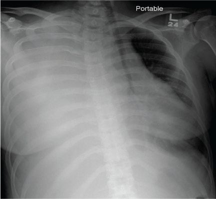
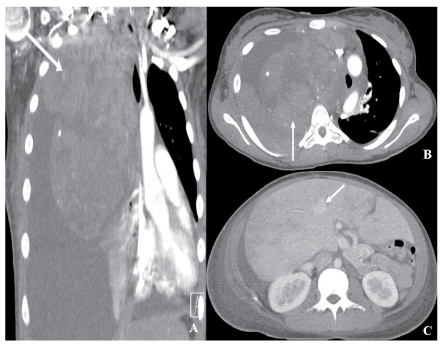
| Initial portal AP chest radiograph (Figure 1) was obtained showed diffuse opacification of the right hemithorax with associated contralateral cardiomediastinal deviation. In addition, there is a suggestion of and slight splaying of the upper right posterior ribs. A follow up IV contrast enhanced Computed Tomography (CT) scan of the chest abdomen and pelvis ensued (Figure 2) to further evaluate findings on the chest radiograph and clinical findings. There was a large, heterogeneous mass with lobulated margins which appeared to be centered in the upper right posterior mediastinum. There was a large associated right pleural effusion and significant mediastinal adenopathy. As suggested on the preceding chest radiograph, there was mass effect with contralateral deviation, compression of the heart and great vessels, tension physiology, and SVC syndrome. Numerous collaterals were seen within the upper anterior and posterior thoracic walls extending to the right supraclavicular region. There was adjacent focal skin and muscle thickening below the axilla near the peripheral aspect of the tumor, which likely represented extrathoracic tumor extension. |
| After initial stabilization, laboratory findings revealed slight microcytic anemia with hemoglobin ranging 10.6-12.0 g/dL (normal range, 12.0-16.0 g/dL) with mean corpuscular volume 77.1 fL (normal range, 79-98 fL), leukocytosis with a white blood cell count of 14.5 ×10³/ µL (normal range, 4.3-11.0×10³/µL) and high neutrophil differential of 90.7% (normal range, 39-77%), and thrombocytosis with platelet count of 953×103/µL (normal range, 140-440×103/µL). Liver enzyme levels were elevated with aspartate aminotransferase level of 91 U/L (normal range, 15-46 U/L) and an alkaline phosphatase level of 177 U/L (normal range, 38-126 U/L). Coagulation studies were abnormal with increased prothrombin time 16.6 secs (normal range, 10.1-12.6 secs). Other abnormal laboratory values include low prealbumin 9 mg/dL (normal range, 20-40 mg/dL), low albumin 2.6 g/dL (normal range, 3.9-5.0 g/dL), elevated lactate dehydrogenase level greater than 2150 U/L (normal range, 313-618 U/L), elevated homovanillic acid 7.7 mg/g creatinine (normal range, 1.4-5.3 mg/g creatinine) and elevated vanillylmandelic acid 4.9 mg/g creatinine (normal range, 1.1-4.1 mg/g creatinine). |
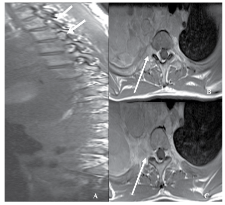
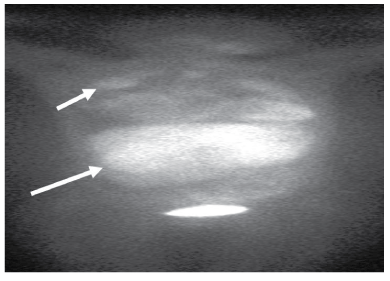
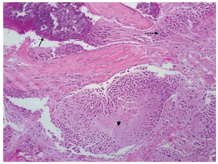
| A contrast-enhanced MRI of the thoracic spine was obtained due to the patients corresponding neurological abnormalities referable to the right upper extremity as well as backpain. (15 ml Multihance was used). This showed that the mass was invading the right upper thoracic neural foramina at the levels of T3-T4 with insinuation along the right dorsolateral epidural space. There was minimal impression on the thecal sac and abnormal marrow signal, which was suspicious for osseous extension of the tumor (Figure 3). Metaiodobenzylguanidine (mIBG) scan (Figure 4) was performed due to the elevated homovanillic and vanillymandelic acid and revealed bilateral lung involvement; right much more than left and also liver and bilateral posterior abdomen involvement. Biopsy of the mass was performed and neuroblastoma confirmed at pathology with neuroblastoma antigen staining (Figures 5-7). |
| Discussion |
| Neuroblastoma, malignant embryonal tumor of the neural crest arising from any part of the sympathetic nervous system, remains the third most common childhood malignancy, after leukemia and CNS tumors. It represents 8-10% of childhood cancers with an incidence per year of 10.5 million children who are less than 15 years of age, and causing 15% of all childhood cancer deaths [1-3]. The median age of presentation overall is 23 months and 40% occur during infancy, 89% by age 5, and 98% by age 10 [4]. It can uncommonly present in adolescence with less than 0.3 cases diagnosed per million people per year, and rarely even well into adulthood, with cases reported even up to 75 years of age [4-6]. The gender ratio of 1:1 in most studies with some showing boys with slightly more frequent than girls at 1.2:1 [1,4,7]. Some risk factors that have been described include maternal alcohol consumption, paternal exposure to nonvolatile and volatile hydrocarbons, diuretic use, pain medications or codeine, low birth weight, and ALK and PHOX2B mutation in familial neuroblastomacases [4,8,9]. These tumors are of ganglion cell origin and are derived from primordial neural crest cells. Consequently, the tumors are found along the typical distribution of sympathetic ganglia, most commonly in the adrenal medulla (35%), but can also be found in the extra-adrenal paraspinal ganglia (30%-35%), followed by the posterior mediastinum in 20%. Interestingly, the disease distribution in younger patients (<1 year) compared to older (>1 year) patients in a previous investigation, found that the mediastinum was an even less common site of presentation in older patients. Other more uncommon sites include the pelvis (2%-3%) and the neck (1%-5%) [2]. |
| The imaging findings of Neuroblastoma are variable, but often well characterized on CT and MRI versus other imaging modalities [10]. Ultrasound shows an echogenic mass with shadowing if calcifications are present [11]. CT is the most commonly used imaging modality for initial assessment and preliminary diagnosis of Neuroblastomas, most often with examination of the chest, abdomen, and pelvis. However, MRI is superior with respect to organ of origin, and regional invasion due to its superior tissue discrimination. Importantly, CT demonstrates the size and extent of the tumor, as well as enhancement characteristics, calcifications, invasion or encasement of vessels and other organs, as well as metastatic lesions [12]. The tumors are often large, lobulated, and heterogeneous in appearance, with variable calcifications and mild, if any enhancement after contrast administration [12]. There may also be hypoattenuating areas of necrosis, or areas of hemorrhage with variable attenuation. Involvement or compression of adjacent structures is common. Invasion of the neural foramina and extension to the epidural space is also common, and can be seen to an extent on CT, but is optimally characterized on MRI. Liver metastases may be diffusely infiltrative, causing global hyperattenuation, or can be in the form of focal hypo enhancing lesions, as in our patient. |
| On MRI, the tumors are heterogenous enhancing masses, with a iso- or hypointense to surrounding tissue on T1 and a hyperintense on T2-weighted fat-suppressed images [12,13]. Signal voids can be seen in areas of calcification. In addition to being the superior modality for organ of origin, it is also the preferred modality for evaluation of neural foraminal and nerve root invasion or displacement, which occurs in 10% of cases, including that of our patient. Bone marrow disease appears as confluent high T2 and low T1 signal within marrow. |
| Another imaging modality used utilizes the function of the neuroendocrine nature of these tumors. Meta-iodobenzylguanidine (mIBG), an analog of norepinephrine, is taken up by catecholamine producing cells and neuroendocrine tumors making radiolabeled mIBG uptake quite specific for neuroblastoma, taken up by 90% to 95% of all neuroblastomas, especially in the pediatric population [12,14,15]. |
| Prognosis is determined by stratification of patients into low-, intermediate-, or high-risk categories based on the Children Oncology Group studies which includes age at diagnosis, INSS stage [1,16], tumor hisopathology, DNA index (ploidy), and MYCN amplification status [17]. Evan’s stage is another prognostic factor. Generally, older ages of presentation, advanced stages (3-4), and presence of cytogenetic abnormalities such as MYCN amplification (25-35%), DNA ploidy, and loss of chromosome 1 p (30-40%) are associated with a poorer prognosis. Neuroblastoma in adolescents and adults appears to differ from that seen in early childhood in several important aspects, as suggested in recent studies. For example, the overall survival appears to be significantly worse and the disease course more indolent. Franks et al. studied a group of 16 such patients over a 27 year period. Overall survival of this group was only 30% at 5 years, whereas the typical overall survival of younger children <1 year is often >70% and can be well into the 90% range with favorable histopathology and single copy MYCN profile [6]. |
| Interestingly, NMYC amplification, a cytogenetic characteristic which is known to be associated with a more aggressive phenotype, was not observed in any of the six patients who were tested. Yet, relapses were observed in all but one, and all but two went on to succumb to their disease. Also, only 40% of those studied had elevations of urinary catecholamines, compared with that expected in this disease, 90-95%. Other studies have also shown poorer survival, more indolent course, and higher morbidity in older patients [7]. Thus, there likely are important biochemical and histopathological differences between Neuroblastomas seen in this population. Treatment includes surgical excision, radiotherapy and chemotherapy depending on risk group classification from the Children Oncology Group studies. Adult cases are rare and are often treated with pediatric guidelines [5]. |
| Given the imaging findings of a low attenuation, lobulated, and mildly enhancing posterior mediastinal mass with scattered calcifications in our 22 years old female, the differential diagnosis would be led by lymphoma. However, the mass in our patient appeared to be centered at the posterior mediastinum, which made lymphoma, a predominantly anterior mediastinal mass, much less likely. Moreover, Mediastinal lymphoma usually is a result of systemic lymphomas (Hodgkin and non-Hodgkin lymphoma) and rarely is the lymphoma isolated to the mediastinum. Hence patients with mediastinal lymphoma generally present with systemic manifestations most commonly the presence of constitutional or “B” symptoms, which were notably absent in our patient [18]. The laboratory findings of elevated urinary HVA and VMA in a relatively young patient also highly favor the diagnosis of Neuroblastoma rather than lymphoma. |
| Other entities such as Osteosarcoma, Metastases, Ewings Sarcoma, and Germ cell tumors would also need to be considered and excluded. In this case, the clinical absence of “B” symptoms and elevated urinary HVA and VMA, favors the diagnosis of Neuroblastoma rather than lymphoma. A metastatic lesion in this setting is also unlikely, in view of the patients age. Furthermore, there was no expansile, enhancing, calcified bone mass suspicious for the rare intra-thoracic osteosarcoma, nor was there a destructive chest wall mass concerning for Ewings Sarcoma. In addition, each of the above differential considerations have associated multimodality imaging findings which were not wholly demonstrated in this case, lending further confidence in the imaging diagnosis of Adult Neuroblastoma, rather than these. |
| The imaging findings of Neuroblastoma can be suggestive, but are not pathognomonic, with some features shared with other entities in the differential diagnosis. Examples of these shared findings can include Low T1 signal or hyperintense signal on T2 weighted imaging, which can also be seen with Lymphoma and Ewing Sarcoma. Neuroblastoma can also be rather heterogenous on CT imaging, which can also be seen with lymphoma. Therefore, the imaging features are best considered along with clinical and laboratory data. MIBG scan is the only exception to this, as it is highly specific for Neuroblastoma. Additional multimodality imaging findings regarding the differential considerations for mediastinal Adult Neuroblastoma include: Low T1 signal intensity with enhancement post-contrast and high T2 signal, with lobulated and complex vascular appearance on ultrasound for Lymphoma, T1 iso- to hypointensity with T2 hyperintensity to skeletal muscle with well-circumscribed soft tissue mass containing variable necrosis, mineralization, and hemorrhage on CT with Osteosarcoma, Variable CT, MRI, and Ultrasound findings in the case of metastasis, and low to intermediate T1 and intermediate to high T2 signal intensity to bone marrow with heterogeneous post-contrast enhancement on CT in the case of Ewing Sarcoma [19-23]. |
| Mediastinal germ cell tumors are rare, and represent less than 5% of all germ cell tumors and with anterior mediastinal location being the most common extragonadal site, representing 50-70% of extragonadal germ cell tumor [24]. Mature teratomas and even less commonly, seminomas are the most frequently seen germ cell tumors in the mediastinum [25]. Occasionally, a mature teratoma may appear in the posterior mediastinum, but only represents up to 8% of all cases [26]. Nonetheless, the majority of mediastinal mature teratoma and 30% of mediastinal seminomas are asymptomatic with symptoms related to the size and location of the tumor and resultant compression of adjacent anatomical structures. [25]. On imaging, they are usually cystic lesions that demonstrate a combination of fat, calcification, fluid and soft tissue attenuation values [27]. These features are absent in our patient’s case. The mediastinum is an uncommon site of presentation compared to the abdomen and pelvis at any age, and is surpassed in rarity only by the cervical region in this respect. |
| In conclusion, our adult patient presented in dramatic fashion with a mediastinal Neuroblastoma, which is known to be a relatively uncommon location for this tumor in patients of any age, but particularly in this age group. Lymphoma would generally lead the differential in this age group from an imaging perspective, however Adult Neuroblastoma may also be considered. Given the considerably worse prognosis of Neuroblastoma in older patients, a high index of suspicion is necessary to avoid unnecessary delays in the diagnosis of this highly uncommon, yet grave variety of this tumor. |
References |
|
Tables and Figures at a glance
| Table 1 | Table 2 |
Figures at a glance
 |
 |
 |
 |
| Figure 1 | Figure 2 | Figure 3 | Figure 4 |
 |
 |
 |
|
| Figure 5 | Figure 6 | Figure 7 |
Post your comment
Relevant Topics
- Abdominal Radiology
- AI in Radiology
- Breast Imaging
- Cardiovascular Radiology
- Chest Radiology
- Clinical Radiology
- CT Imaging
- Diagnostic Radiology
- Emergency Radiology
- Fluoroscopy Radiology
- General Radiology
- Genitourinary Radiology
- Interventional Radiology Techniques
- Mammography
- Minimal Invasive surgery
- Musculoskeletal Radiology
- Neuroradiology
- Neuroradiology Advances
- Oral and Maxillofacial Radiology
- Radiography
- Radiology Imaging
- Surgical Radiology
- Tele Radiology
- Therapeutic Radiology
Recommended Journals
Article Tools
Article Usage
- Total views: 14933
- [From(publication date):
July-2013 - Jul 02, 2025] - Breakdown by view type
- HTML page views : 10350
- PDF downloads : 4583
