Mini Review Open Access
New Advances in Three-Dimensional Endoscopic Surgery
Kenoki Ohuchida1,2, Nagai Eishi2, Satoshi Ieiri3, Akahoshi Tomohiko3, Ikeda Tetsuo3, Masao Tanaka2 and Makoto Hashizume1,3*1Department of Advanced Medical Initiatives, Graduate School of Medical Sciences, Kyushu University, Fukuoka, Japan
2Department of Surgery and Oncology, Graduate School of Medical Sciences, Kyushu University, Fukuoka, Japan
3Department of Advanced Medicine and Innovative Technology, Kyushu University Hospital, Fukuoka, Japan
- *Corresponding Author:
- Makoto Hashizume
Department of Advanced Medical Initiatives
Faculty of Medical Sciences, Kyushu University
3-1-1 Maidashi, Fukuoka 812-8582, Japan
Tel: +81-92-642-5993
Fax: +81-92-642-5991
E-mail: mhashi@dem.med.kyushu-u.ac.jp
Received date: October 14, 2013; Accepted date: November 18, 2013; Published date: November 26, 2013
Citation: Ohuchida K, Eishi N, Ieiri S, Tomohiko A, Tetsuo I (2013) New Advances in Three-Dimensional Endoscopic Surgery. J Gastroint Dig Syst 3:152. doi: 10.4172/2161-069X.1000152
Copyright: © 2013 Ohuchida K, et al. This is an open-access article distributed under the terms of the Creative Commons Attribution License, which permits unrestricted use, distribution, and reproduction in any medium, provided the original author and source are credited.
Visit for more related articles at Journal of Gastrointestinal & Digestive System
Abstract
The indications for minimally invasive surgery continue to expand worldwide, with endoscopic surgery currently regarded as standard treatment in many surgical fields. The prototype of a three-dimensional [3D] system for endoscopic surgery was developed during the early 1990s to overcome the lack of depth perception on a twodimensional [2D] display. The prototype, however, was not widely used because of the poor quality of the image and visual side effects. Advances in the technology have now allowed the development of 3D endoscopic systems with high-definition resolution that are commercially available. We discuss the development, basic studies, present status, and future of 3D endoscopic systems, including their advantages and disadvantages. Although further improvements are needed for more advanced surgery, the rapid advances being experienced in 3D endoscopic system will hopefully overcome the remaining problems quickly and provide the ultimate version of minimally invasive surgery.
Keywords
Three-dimensional surgery; Stereoscopic surgery; 3D
Lack of Depth Perception in Conventional Endoscopic Surgery
The indications for minimally invasive surgery continue to expand. In fact, endoscopic surgery is currently regarded as standard treatment in numerous surgical endeavors. Advances in endoscopic surgery have provided patients with less postoperative pain, shorter hospital stays, and an earlier return to work. For the surgeon, however, endoscopic surgery seems to be more stressful than open surgery. For example, several skills that are not required for open surgery are necessary for endoscopic surgery. The lack of stereoscopic vision on a twodimensional [2D] display is one of the most significant difficulties for the endoscopic surgeon, who performs laparoscopic surgery or thoracoscopic surgery. The lack of stereoscopic vision on a 2D display causes visual misperceptions based on the loss of depth perception. To compensate for this lack, expert endoscopic surgeons use visual clues from a 2D display, such as object interposition, relative motion of the endoscope, familiar anatomy, and the size of anatomical structures [1]. Acquiring familiarity with, and the ease of identifying, these landmarks takes an investment of much time and effort. Thus, the endoscopic surgeon must invest a great deal of time to gain the extensive experience necessary to become familiar with these depth clues and overcome the lack of depth perception.
Developments and Basic Study of the Three-Dimensional Endoscope and Display
The three-dimensional [3D] system for endoscopic surgery was developed during the early 1990s to overcome the lack of depth perception on a 2D display. Becker et al. [2] and other researchers [3-7] investigated the effects of 3D endoscopes and displays on endoscopic procedures. Whereas several of these studies reported significant benefits of these 3D systems [4,7,8], others did not [3,6]. When the prototype of the 3D system was first used for endoscopic surgery, the surgeons themselves sometimes developed side effects such as headaches, ocular fatigue, and dizziness caused by the heavy active shutter glasses and the poor quality in the 3D images [3]. Another roadblock to its use was the high purchase cost, which kept it from becoming commercially available. Hence, the earlier 3D systems were not widely used.
Researchers have now reported some advances in 3D endoscopic systems [9-11]. Birkett [11] reported greater comfort with the new light-polarizing glasses compared to the earlier active shutter glasses. Yamauchi and Shinohara [10] used a current stereoscopic endoscope modified to match the relation between binocular disparity and convergence. They compared fatigue during endoscopic procedures under 2D and 3D visual conditions and found no differences between them.
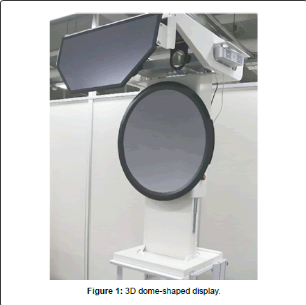
We also have developed a novel 3D dome-shaped display [3DD] system with high resolution [XGA] and designated it Cyber Dome (Figure 1) [12]. Our purpose was to provide more depth perception for endoscopic surgeons than what had been provided with 3D endoscopic systems using a plain display [3DP]. In addition, our 3DD system does not require a shutter mechanism in the glasses, which often causes significant side effects as well as dark images when using the prototype of the 3DP system. To aid in evaluating the efficacy of the Cyber Dome, we designed and applied six new tasks as well as the conventional tasks of suturing and knot tying. We evaluated the effects of the novel 3DD system on endoscopic surgery and compared the results with those attained using a conventional 2D system, with particular focus on depth perception. We found that the 3DD system significantly improved depth perception based on its ability to perform the six new tasks. Its use also reduced the execution time and the number of errors in suturing and knot tying.
Present Status of 3D Endoscopic Systems
HD systems are often used in conventional 2D endoscopic systems to provide extremely high resolution for the surgeon. Currently, however, 3D endoscopic systems with HD resolution have become commercially available from several vendors (Table 1), including Shinko Optical Co. [Tokyo, Japan, http://www.shinko-koki.jp/ shi3d_02_01.html], Olympus Co. [Tokyo, Japan, http://www.olympusglobal.com/en/news/2013a/nr1304093dscopee.jsp], KARL STORZ [Tuttlingen, Germany, https://www.karlstorz.com/cps/rde/xchg/SID-56FE1E72-66DF5BD6/karlstorz-en/hs.xsl/16957.htm], and Viking Systems [Westborough, MA, US, http://www.ncbi.nlm.nih.gov/pmc/articles/PMC3349929/]. Shinko Optical, and Viking Systems provide a rigid, angulated, dual-channel rod lens laparoscope with two camera sensors. Olympus provides an articulating HD 3D laparoscopic surgical video system. Usually, such a 3D system consists of the display with a shutter system that delivers images to the right and left eyes by sequential presentation on a single display. The surgeon using this system is required to wear circular polarized glasses. Also available now is a dome-shaped display on which two projectors deliver images to the right and left eyes, respectively, via simultaneous presentation. This product is available from Panasonic Healthcare Co. Ltd. (Osaka, Japan).
| Company | 3D Scope | Function |
| Olympus | Flexible type 10 mm | HD system Articulating scope |
| Shinko Optical | 0, 30 degrees rigid type 5 mm, 10mm | 3CCCD HD system Giro function |
| KARL STORZ | 0, 30 degrees rigid type 10 mm | HD system |
| Viking Systems | 0, 30 degrees rigid type 10 mm | HD system |
Table 1: Comparison of 3D Systems.
Clinical Experience of the use of 3D Endoscopic Systems
During the 1990s, a prototype 3D endoscopic system was introduced for use in laparoscopic surgery. At that time, only simple procedures, such as cholecystectomy, were performed with 3D laparoscopic surgery. 3D images for simple endoscopic procedures may not improve the skills of experienced laparoscopic surgeons because these surgeons do not require depth perception to perform simple procedures. 3D images may be more useful for complicated procedures, such as advanced laparoscopic surgery on a cancerous growth.
It is hopefully expected that endoscopic surgery can provide not only a less invasive approach but can contribute to better results with more sophisticated procedures and more accurate lymph node dissection during advanced surgery for cancer than can be achieved by open surgery. One of the reasons for this assertion is that it provides an enlarged view during endoscopic surgery.
Endoscopic systems that support full HD 2D video have been introduced in recent years. Such a high-quality 2D endoscopic system allows endoscopic surgeons to observe the fine structures of tissue and organs that are not visible to the naked eye during conventional open surgery. Hence, endoscopic surgery is expected to be not only less invasive but also more suitable than conventional open surgery when more precision is required [e.g., surgery requiring lymph node dissection]. The new 3D endoscopic systems, which provide HD images, have already been used for laparoscopic surgery to remove gastric, colorectal, pancreatic, and hepatic lesions.
Experienced endoscopic surgeons can use shadow or movement parallax as depth cues instead of stereovision. Therefore, they may not always need a 3D system. It has been reported that 97% of surgical accidents during laparoscopic cholecystectomy occur as a result of visual misperceptions [13]. We reported that the 3D system significantly reduced the number of errors incurred when using endoscopic forceps [12]. Hofmeister et al. [14] and Chan et al. [3] reported that 2D display systems can cause optical illusions, and thus erroneous maneuvers. Taken together, these findings suggest that the 3D endoscopic system may help reduce the number of surgical accidents. It can also be useful for advanced laparoscopic surgery such as fine lymph node dissection and complicated reconstruction after resection. The 3D system provides useful information via depth perception even to expert surgeons performing these complicated procedures.
Since 2009, we have routinely used a 3D endoscopic system in more than 200 cases including gastric, pancreatic, pediatric, colorectal, urologic, gynecologic, and thoracoscopic surgery as well as for splenectomy, brain surgery, and endlaryngeal microsurgery. We used three 3D systems that had been developed by Shinko Optical Co., Olympus Co., and Karl Storz GmbH & Co, respectively. We also used 5.4-and 4.7-mm 3D endoscopes and 3-mm3D endoscopes [Shinko Optical Co.] for brain surgery and for endolaryngeal microsurgery.
Benefits of 3D Endoscopic Systems
Lymph node dissection
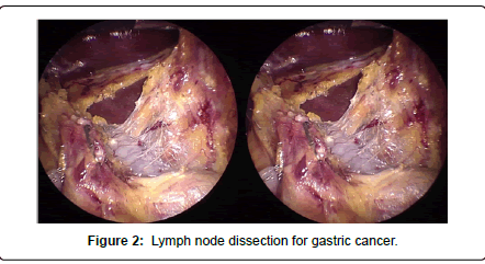
Surgeons can perform fine lymph node dissection with oncological safety using a 2D endoscopic system during advanced laparoscopic surgery for cancer. An enlarged view of the 2D laparoscope gives surgeons detailed information of anatomical tissue structures and contributes to the surgeon’s decision of suitable resection lines to maintain oncological safety. A 3D endoscopic system provides the same benefits to laparoscopic surgeons but with depth perception. Information about the depth is especially useful for making intuitive decisions about the direction of the traction of tissues. Thus, it is now possible for surgeons to move an instrument in the suitable direction with confidence. For example, even if the target tissue is fragile, the surgeon can hold it in a suitable direction without damaging it, maintaining the appropriate tension to still have the large surgical field of view (Figure 2).
Reconstruction of the digestive tract
Advanced laparoscopic surgery can accomplish intracorporeal reconstruction of the digestive tract with operations such as gastroduodenostomy, gastrojejunostomy, and esophagojejunostomy. Surgeons often use linear staplers during these procedures, so it is important to determine the direction of the remnant stomach, small intestine, or esophagus. Because the operator performs these procedures in cooperation with an assistant, each needs to know the direction of the linear staplers and the organs. With a 2D endoscopic system, “these partners” compensate for the lack of the information regarding the direction based on their experience, but usually their experiences have not been precisely the same. With the 3D endoscopic system, they can take advantage of having the same information regarding depth derived from the 3D image and can perform the procedures with confidence. This situation may lead to increased accuracy and patient safety.
Future of 3D Endoscopic Systems
Several 3D endoscopic systems that provide much higher resolution than previous 3D prototypes are commercially available. These systems do not induce the adverse visual symptoms-nausea, visual fatigue, visual disturbance-that were experienced with the previous 3D prototypes. The quality of the current 3D image is relatively low, however, when compared with the 3D system incorporated into the robotic system that is often used today. Currently, a master/slave-type surgical robotic system is used to perform robotic surgery. The only commercially available robotic system today is the da Vinci® Surgical System [Surgical Intuitive], which provides visualization through a high-quality 3D endoscope and has been used mainly for prostatectomy or histerectomy. The newest da Vinci® Surgical System models have a fully HD 3D video system, which provides depth perception for surgeons as well as HD images. It contributes to increased safety during procedures and improves accuracy during lymph node dissection, which in turn would increase curability associated with resection of malignancies. Although the da Vinci® Surgical System 3D system provides high-quality 3D images, the endoscope is too large and too heavy to hold by the human hand during the surgery-thereby necessitating the use of a robotic arm. In contrast, the size and weight of nonrobotic 3D endoscopes in current 3D endoscopic systems are similar to the conventional 2D endoscope and are easy to hold in the human hand during surgery.
A new model of the 3D endoscopic system with 4K CCD chips is under development. This new system will provide full HD 3D imagesthe same as the da Vinci® surgical system. Such images will contribute to the accuracy and safety of the procedures during complicated advanced surgery, such as oncological surgery with fine lymph node dissection.
Conclusion
The 3D endoscopic systems have both advantages and disadvantages. The further development of technologies and equipment is necessary to allow safe, widespread introduction of 3D endoscopic systems for use during advanced surgery, especially for malignant tumors. Although the initial cost of a 3D endoscopic system represents a large investment, more widespread use these 3D endoscopic systems may resolve this problem. Despite several problems with its use, the 3D endoscopic system has already been used in numerous surgical fields. Advances in 3D endoscopic system will hopefully overcome these problems and provide the ultimate version of minimally invasive surgery.
Acknowledgement
Supported in part by a grant from the New Energy and Industrial Technology Development Organization.
References
- Byrn JC, Schluender S, Divino CM, Conrad J, Gurland B, et al. (2007) Three-dimensional imaging improves surgical performance for both novice and experienced operators using the da Vinci Robot System. Am J Surg 193: 519-522.
- Becker H, Melzer A, Schurr MO, Buess G (1993) 3-D video techniques in endoscopic surgery. Endosc Surg Allied Technol 1: 40-46.
- Chan AC, Chung SC, Yim AP, Lau JY, Ng EK, et al. (1997) Comparison of two-dimensional vs three-dimensional camera systems in laparoscopic surgery. Surg Endosc 11: 438-440.
- Dion YM, Gaillard F (1997) Visual integration of data and basic motor skills under laparoscopy. Influence of 2-D and 3-D video-camera systems. Surg Endosc 11: 995-1000.
- van Bergen P, Kunert W, Buess GF (2000) The effect of high-definition imaging on surgical task efficiency in minimally invasive surgery: an experimental comparison between three-dimensional imaging and direct vision through a stereoscopic TEM rectoscope. Surg Endosc 14: 71-74.
- Hanna GB, Shimi SM, Cuschieri A (1998) Randomised study of influence of two-dimensional versus three-dimensional imaging on performance of laparoscopic cholecystectomy. Lancet 351: 248-251.
- Wenzl R, Lehner R, Vry U, Pateisky N, Sevelda P, et al. (1994) Three-dimensional video-endoscopy: clinical use in gynaecological laparoscopy. Lancet 344: 1621-1622.
- van Bergen P, Kunert W, Bessell J, Buess GF (1998) Comparative study of two-dimensional and three-dimensional vision systems for minimally invasive surgery. Surg Endosc 12: 948-954.
- Taffinder N, Smith SG, Huber J, Russell RC, Darzi A (1999) The effect of a second-generation 3D endoscope on the laparoscopic precision of novices and experienced surgeons. Surg Endosc 13: 1087-1092.
- Yamauchi Y, Shinohara K (2005) Effect of binocular stereopsis on surgical manipulation performance and fatigue when using a stereoscopic endoscope. Stud Health Technol Inform 111: 611-614.
- Birkett DH (1994) Three-dimensional laparoscopy in gastrointestinal surgery. Int Surg 79: 357-360.
- Ohuchida K, Kenmotsu H, Yamamoto A, Sawada K, Hayami T, et al. (2009) The effect of CyberDome, a novel 3-dimensional dome-shaped display system, on laparoscopic procedures. Int J Comput Assist Radiol Surg 4: 125-132.
- Way LW, Stewart L, Gantert W, Liu K, Lee CM, et al. (2003) Causes and prevention of laparoscopic bile duct injuries: analysis of 252 cases from a human factors and cognitive psychology perspective. Ann Surg 237: 460-469.
- Hofmeister J, Frank TG, Cuschieri A, Wade NJ (2001) Perceptual aspects of two-dimensional and stereoscopic display techniques in endoscopic surgery: review and current problems. Semin Laparosc Surg 8: 12-24.
Relevant Topics
- Constipation
- Digestive Enzymes
- Endoscopy
- Epigastric Pain
- Gall Bladder
- Gastric Cancer
- Gastrointestinal Bleeding
- Gastrointestinal Hormones
- Gastrointestinal Infections
- Gastrointestinal Inflammation
- Gastrointestinal Pathology
- Gastrointestinal Pharmacology
- Gastrointestinal Radiology
- Gastrointestinal Surgery
- Gastrointestinal Tuberculosis
- GIST Sarcoma
- Intestinal Blockage
- Pancreas
- Salivary Glands
- Stomach Bloating
- Stomach Cramps
- Stomach Disorders
- Stomach Ulcer
Recommended Journals
Article Tools
Article Usage
- Total views: 16790
- [From(publication date):
November-2013 - Nov 16, 2025] - Breakdown by view type
- HTML page views : 11957
- PDF downloads : 4833


