Research Article Open Access
Identification of Hylemonella gracilis as an Antagonist of Yersinia pestis Persistence
David R Pawlowski1,2*, Amy Raslawsky2, Gretchen Siebert2, Daniel J Metzger2, Gerald B Koudelka3 and Richard J Karalus1,2
2Department of Microbiology and Immunology, School of Medicine, State University of New York at Buffalo
3Department of Biological Sciences, College of Arts and Sciences, State University of New York at Buffalo
- *Corresponding Author:
- David R Pawlowski
CUBRC, Inc., State University of New York at Buffalo
716-829-2236, 3435 Main Street
134 Biomedical Research Building
Buffalo, NY 14214, USA
E-mail: Pawlowski@cubrc.org, drp@buffalo.edu
Received Date: October 14, 2011; Accepted Date: December 13, 2011; Published Date: December 18, 2011
Citation: Pawlowsk DR, Raslawsky A, Siebert G, Metzger DJ , Koudelka GB, et al. (2011) Identification of Hylemonella gracilis as an Antagonist of Yersinia pestis Persistence. J Bioterr Biodef S3:004. doi: 10.4172/2157-2526.S3-004
Copyright: © 2011 Pawlowsk DR, et al. This is an open-access article distributed under the terms of the Creative Commons Attribution License, which permits unrestricted use, distribution, and reproduction in any medium, provided the original author and source are credited.
Visit for more related articles at Journal of Bioterrorism & Biodefense
Abstract
Yersinia pestis , the etiological agent of plague, has garnered great interest in the Biological Defense community as its intentional release or use as a terror weapon could cause considerable morbidity while incurring incalculable financial costs for restoration and remediation efforts. The plague bacterium is thought to only persist within a host such as a flea or small mammal reservoir. Following an event such as an intentional release however, the plague bacterium would be spread throughout a number of atypical environments such as soil or water ecosystems. Recently, a small number of studies have been published describing the plague bacterium’s persistence in some of these atypical environments. Here we show that Y. pestis can colonize sterilized water microcosms for over 3 years yet when introduced to filtered fresh water microcosms the bacterium Hylemonella gracilis became the dominant bacterium in the microcosm, apparently preventing long-term Y. pestis persistence. The conditioning and outgrowth of H. gracilis on rich media is directly attributable and proportional to the introduction and concentration of Y. pestis to the microcosm.
Introduction
Yersinia pestis, the etiological agent of the plague, is reported to be responsible for three pandemics throughout recorded history [1]. The plague manifests a number of disease states in infected individuals including bubonic, pneumonic, systemic and enteropathogenic modalities [2,3]. The high morbidity and mortality rates associated with the plague have made it a Category A pathogen with a relatively high potential for use as a bioterror weapon [4]. In the event of an intentional release of Y. pestis, the plague bacterium could be spread throughout a wide variety of environments ranging from natural to manmade. A complete understanding of the plague bacterium’s fate upon exposure to these environments would enhance the ability of first responders, policy makers, and those involved in restoration efforts how best to apply limited resources.
In another sense, an understanding of the fate of Y. pestis in the environment could help to explain gaps in the classical model of plague maintenance and disease [5,6]. The classical model identifies small mammals as the reservoir accompanied by flea-mediated transmission. While this model fits typical transmission patterns during epizootic cycles, plague maintenance during enzootic periods is poorly understood. Recently, a small number of reports attempting to delineate Y. pestis’ persistence during enzootic periods have appeared [5,7,8]. These reports suggest alternative plague life-cycle possibilities in the context of a host or vector. Other reports suggest that telluric plague may aid in Y. pestis persistence [9,10]. Historical data shows that viable Y. pestis can be found in the soil. For example, Yersin first isolated Y. pestis from the soil floor of a house where an infection had occurred [11]. In another example, Mollaret had demonstrated that telluric plague maintained pathogenicity for approximately 40 weeks [12]. These results were verified decades later by Ayyadurai et al. [13]. Taken together, these reports and associated data indicate terrestrial environments may play a role in Y. pestis’ maintenance during enzootic periods.
As Y. pestis studies progress, it is becoming increasingly clear that Y. pestis can survive outside of a host. We wish to better understand the types of terrestrial environments tolerated by Y. pestis and to gain insight into factors that may influence survivability in those environments. Our recently published work [14] and that of others [15], indicated that Y. pestis can survive in fresh water. One unexpected result was the discovery that Y. pestis can enter a dormant or viable but non-culturable state when inoculated into a tap water microcosm [14]. These findings imply that freshwater may provide a reservoir for Y. pestis in the environment and that the persistence of plague bacterium could impact risk assessment and remediation efforts, especially in the event of an intentional release of this pathogen. To extend our work on the ability of Y. pestis to survive in atypical environments we found that the plague can survive in autoclaved river water for over three years. This persistence study also revealed that a common fresh water bacterium, Hylemonella gracilis was stimulated for growth by the addition of Y. pestis that, in turn, appears to have prevented longterm colonization by Y. pestis. Sequence analysis confirmed H. gracilis as the bacterium isolated in these studies. Finally, we showed that the stimulation and amount of H. gracilis outgrowth was specific and proportional to Y. pestis inoculum concentration and sensitive to 50 colony forming units per milliliter.
Information concerning Hylemonella gracilis is limited [16-18]. However, Wang et al. shows that H. gracilis is capable of passing through 0.45μm, 0.22μm, and 0.1μm filters [17]. This observation provides a sound explanation for the persistence of H. gracilis in our filtered river water microcosms.
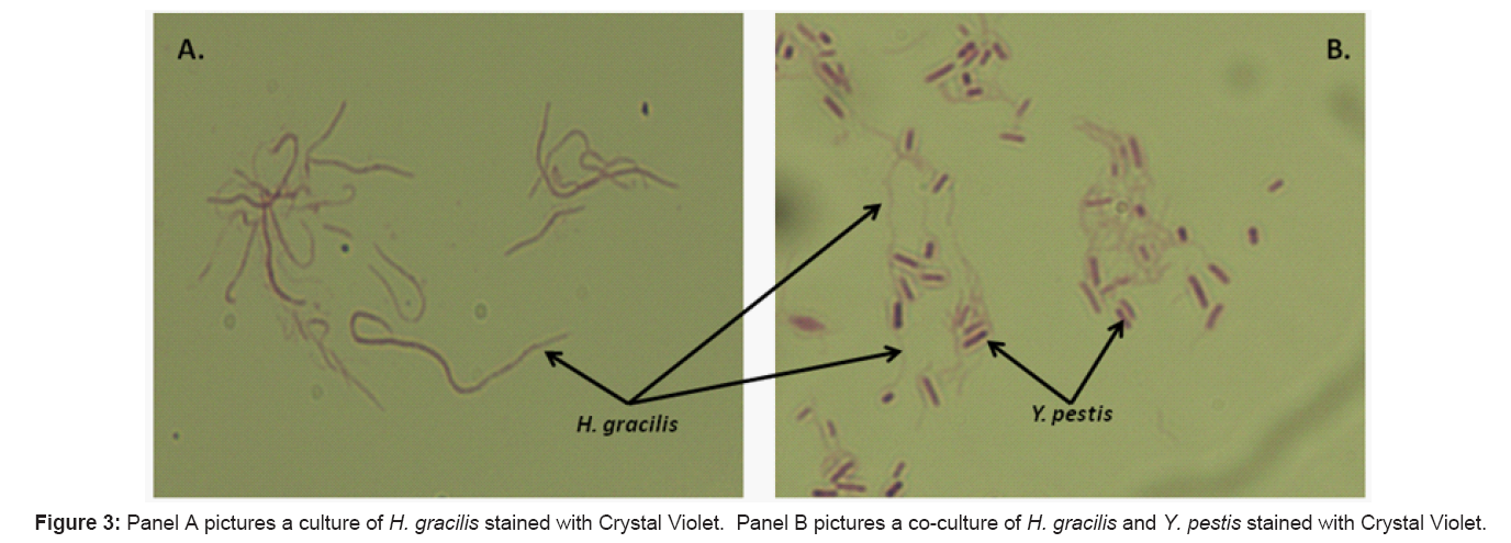
Hylemonella gracilis is classified within the Betaproteobacteria class, Burkholderiales order and Comamondaceae family. This bacterium was previously classified as Aquaspirillum gracile and originally identified as Spirillum gracile, a common microbe to fresh water ecosystems throughout the world [16,19]. Stains of the H. gracilis strain that we isolated display morphologies identical to the historical descriptions (Figure 3) [19,20].
Materials and Methods
Bacterial strains and media
Yersinia pestis strain A1122 was utilized for these experiments. The A1122 strain (Orientalis Biovar) is avirulent, lacking the 103 kb Pgm locus and the Lcr plasmid. Other strains used were Y. pseudotuberculosis (ATCC# 29833) Y. enterocolitica (9610), Y. rohdei (43380), Y. ruckeri (29473), Y. frederiksenii (33641), Y. intermedia (29909) and Escherichia coli (25922). Y. pestis A1122, Y. pseudotuberculosis, and Y. enterocolitica were grown to early stationary phase in Brain Heart Infusion (BHI) broth at 37°C in a shaking water bath.
NRW was collected from the upper Niagara River in mid-August, 2007 and stored at 4°C until needed. Following filter or autoclave sterilization, the water microcosms were determined to be free of colony forming bacteria by plating on multiple agar types (Table 1). NRW was filter sterilized using Corning 1L bottle top 0.22μm cellulose acetate filters.
| Agar (solid) media | Filtered NRW | Filtered NRW + Y.Pestis |
|---|---|---|
| Yeast Peptone Dextrose (YPD) | No growth | No growth |
| Mueller Hinton | No growth | No growth |
| Middlebrook 7H10 | No growth | No growth |
| Luria Bertani (LB) | No growth | No growth |
| Brain Heart Infusion (BHI) | No growth | No growth |
| Mycoplasma | No growth | No growth |
| Nutrient | No growth | Extensive growth |
| Tryptic Soy Agar + 5% Sheep’s blood (TSA II) |
No growth | Moderate growth |
| ATCC Medium 233 Spirillum gracile medium | No growth | Extensive growth |
Table 1: Outgrowth of unknown bacteria from Niagara river water.
Yersinia pestis persistence
The specified number of colony forming units per milliliter (CFU/ mL) was added to triplicate microcosms of either filtered or autoclaved NRW. The bacterial concentration was determined by serial dilution plating on TSA II agar at the times specified in the results section. Incubations were done at room temperature (~26°C)
Identification of H. gracilis
16S rRNA gene sequencing: Three single colonies of H. gracilis grown on TSA II from a river water microcosm inoculated with Y. pestis were suspended in PBS and the 16S rRNA genomic DNA amplified with primer pair set, 27f and 787r, using amplification conditions described by Whitehouse et al. [3]. Following amplification, the PCR products were separated in a 1.2% agarose gel. The amplified DNA was sent to the Roswell Park Cancer Institute’s Biomolecular resources facility, DNA sequencing laboratory for Sanger sequencing. The resulting sequence was queried against the NCBI database using the BLASTn suite.
Simple staining: A colony of each Y. pestis and H. gracilis was resuspended in one milliliter of PBS, vortexed for thirty seconds and incubated at room temperature for 20 minutes. Five microliters of the co-culture was spread on a glass slide and air dried. Simultaneously, a colony of H. gracilis was resuspended in 10 microliters of PBS directly on a slide and dried. The dried cultures were heat-fixed to the slide by passing over an open flame. The slides were flooded with Crystal Violet stain (Difco BBL) for 45 seconds and washed with distilled water. The slides were air dried and visualized by light microscopy using a Nikon Eclipse 80i microscope equipped with an Infinity 3 digital camera (Lumenera Corp., Ottawa, ON).
Yersinia pestis specificity and sensitivity: Single microcosms of filtered NRW were inoculated with Y. pestis to final concentrations of 5 x 101- 5 x 10⁷ CFU/mL. Single microcosms of filtered NRW were also inoculated with 5 x 10³, 5 x 10⁵ and 5 x 10⁷ CFU/mL for each of the other bacteria listed. The number of CFUs/mL of each organism was monitored by triplicate plating on TSA II agar as a function of time at 26oC.
Results
Yersinia pestis persistence
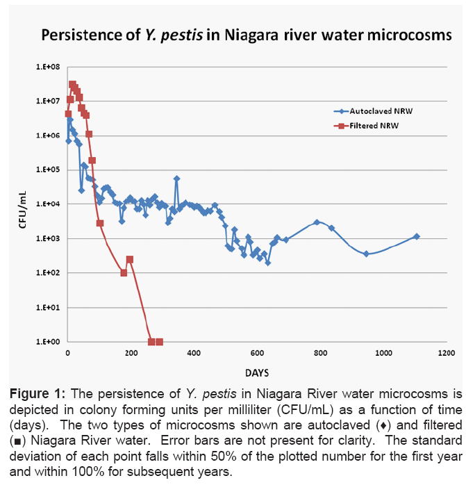
Yersinia pestis A1122 was inoculated into either filtered or autoclaved NRW microcosms to a concentration of approximately 5 x 10⁶ CFU/mL. The persistence of Y. pestis as a function of time was observed by serial dilution plating. The results in Figure 1 show that viable Y. pestis persists in autoclaved Niagara River water for over 3 years. In these microcosms, a die-off of approximately 99% of the original population occurred within 100 days post-inoculation. Following this die-off, the population slowly declined until it reached approximately 0.1% of its initial inoculum concentration at which point the number of Y. pestis stabilized at approximately 500-1000 CFU/mL. These data are in agreement with other long-term bacterial survival studies where phases of growth, decline and maintenance have been identified and characterized [21,22].
Figure 1: The persistence of Y. pestis in Niagara River water microcosms is depicted in colony forming units per milliliter (CFU/mL) as a function of time (days). The two types of microcosms shown are autoclaved (♦) and filtered (■) Niagara River water. Error bars are not present for clarity. The standard deviation of each point falls within 50% of the plotted number for the first year and within 100% for subsequent years.
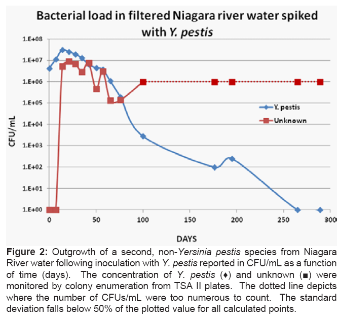
In contrast to the results obtained using autoclaved NRW, the population of Y. pestis dropped to extinction within 265 days in the filtered NRW water microcosm (Figure 1). We also found that the Y. pestis treated filtered NRW microcosm became overrun with a second bacterium. To begin to characterize this phenomenon, we monitored the amount of Y. pestis and this second, non-Y. pestis bacterium (Figure 2). We found that this bacterium increased to 5 x 10⁶ CFU/mL within 14 days post-inoculation of the microcosm with Y. pestis, while the amount of Y. pestis decreased concomitantly. The population of the second bacterium reached a maximum at ~21 days and was maintained within a log of this level for at least 280 days.
Figure 2: Outgrowth of a second, non-Yersinia pestis species from Niagara River water following inoculation with Y. pestis reported in CFU/mL as a function of time (days). The concentration of Y. pestis (♦) and unknown (■) were monitored by colony enumeration from TSA II plates. The dotted line depicts where the number of CFUs/mL were too numerous to count. The standard deviation falls below 50% of the plotted value for all calculated points.
The correlation between growth of the second bacterium and the decrease in Y. pestis led us to hypothesize that an interaction between Y. pestis and this unknown bacterium may cause a decline of the plague bacterium in the filtered NRW microcosm (Figure 2). To begin to test this idea, we generated several replicates of this microcosm using independent cultures of Y. pestis to inoculate filtered NRW in order to rule out contamination from the original culture. The results from the replicate microcosms were consistent with the original indicating that exogenous contamination was not responsible for the outgrowth of the second bacterial species (data not shown). We also tested the filtered river water itself for the presence of the bacterium prior to the addition of Y. pestis. To test for the presence of the second bacterium, we plated samples of filtered NRW on a variety of agar based culture media (Table 1). The results of these tests show that prior to the addition of the Y. pestis there was no bacterial growth. However upon addition of Y. pestis A1122 the unknown bacterium displayed growth on TSA II agar, Nutrient agar and ATCC Media 233: Spirillum gracile medium (Table 1).
Identification of Hylemonella gracilis
To establish the identity of the bacterium that was conditioned for growth on nutrient rich agars by Y. pestis introduction into fresh water microcosms, we determined the sequence of the 16S rRNA of this bacterium. The 16S rRNA sequence was used as a probe of the NCBI’s database. We performed two BLAST queries, one against all sequences and a second against all microbial genomes in the database. The top ten hits obtained from each query are shown in Tables 2 and 3 respectively. These results clearly identify the induced bacteria as Hylemonella gracilis. Microscopic visualization of the bacterium shows a flexible, elongated shape consistent with previous descriptions of H. gracilis (Figure 3, Panels A and B) [19].
| Accession # | Description | Query coverage | Max ID |
|---|---|---|---|
| AJ565423.1 | Hylemonella gracilis partial 16s rRNA gene, isolate WQP1 | 100% | 100% |
| AJ565426.1 | Hylemonella gracilis partial 16s rRNA gene, isolate MWH-Mo30.9.Ko2 | 100% | 100% |
| AJ565425.1 | Hylemonella gracilis partial 16s rRNA gene, isolate WQM2 | 100% | 100% |
| AJ565428.1 | Hylemonella gracilis partial 16s rRNA gene, isolate MWH-Tang4W16 | 100% | 99% |
| AJ565424.1 | Hylemonella gracilis partial 16s rRNA gene, isolate WQT2 | 100% | 99% |
| NR 024934.1 | Hylemonella gracilis strain ATCC 19624 16S ribosomal RNA, partial sequence>gb│AF078753.1│Aquaspirillum gracile 16S ribosomal RNA gene, partial sequence | 100% | 99% |
| AJ565429.1 | Hylemonella gracilis partial 16s rRNA gene, isolate MWH-7W8 | 100% | 99% |
| AJ565427.1 | Hylemonella gracilis partial 16s rRNA gene, isolate MWH-Tang3W12 | 100% | 99% |
| AB539986.1 | Hylemonella gracilis 16s rRNA, partial sequence, strain: IZ9-1 | 97% | 100% |
| AJ565423.1 | Hylemonella gracilis partial 16s rRNA gene, isolate WQP1 | 100% | 100% |
Table 2: Top ten BLAST results from the sequence database query.
| Accession # | Description | Query coverage | Max ID |
|---|---|---|---|
| AEGR01000103.1 | Hylemonella gracilis ATCC 19624 Contig80, whole genome shotgun sequence | 98% | 99% |
| NC 015138.1 | Acidovorax avenae subsp. Avenae ATCC19860 chromosome, complete genome | 98% | 98% |
| NC 008752.1 | Acidovorax citrulli AAC00-1 chromosome, complete genome | 98% | 98% |
| NC 015677.1 | Ramlibactertataouinensis TTB310 chromosome, complete genome | 98% | 98% |
| NC 012791.1 | Variovorax paradoxus S110 chromosome chromosome 1,complete sequence | 98% | 98% |
| NC 010002.1 | Delftia acidovorans SPH-1 chromosome, complete genome | 98% | 98% |
| NC 015563.1 | Delftia sp. Cs1-4 chromosome, complete genome | 98% | 98% |
| NC 015422.1 | Alicycliphilus denitrificans K601 chromosome, complete genome | 98% | 98% |
| NC 014910.1 | Alicycliphilus denitrificans BC chromosome, complete genome | 98% | 98% |
| NC 014931.1 | Variovorax paradoxus EPS chromosome, complete genome | 98% | 98% |
| NC 011992.1 | Acidovorax ebreus TPSY chromosome, complete genome | 98% | 98% |
Table 3: Top ten BLAST results from the bacterial genome database query.
Specificity and sensitivity of H. gracilis for Y. pestis
We next looked at both the specificity and sensitivity of the interaction between H. gracilis and Y. pestis. The specificity of H. gracilis conditioning and outgrowth by Y. pestis addition was monitored by inoculating filtered NRW with a variety of other Yersinia species (Material and Methods) and Escherichia coli. Of these, only Y. enterocolitica induced any outgrowth of H. gracilis indicating a highly specific interaction stimulates the outgrowth of this bacterium.
Sensitivity of the polymicrobial interaction between Y. pestis and H. gracilis was determined by titrating the size of the inoculum. In these experiments, microcosms of filtered NRW were inoculated with tenfold dilutions of Y. pestis, Y. enterocolitica and
Figure 4: The persistence of Y. pestis (♦), Panels A and C, and Y. enterocolitica (♦), Panel B, were determined while simultaneously monitoring for H. gracilis (■) outgrowth. Initial inoculating concentrations were 5 x 10⁶ CFU/mL (Panels A and B), 50 CFU/mL (Panel C), and 500 CFU/mL (Panel D). The dotted line in Panel A depicts where the number of CFUs/mL of H. gracilis were too numerous to count. No standard deviations were calculated as these data were generated from triplicate platings of a single microcosm.
Discussion
The threat posed by the intentional release of biological agents and the lack of information regarding their behavior in non-traditional environments has lead a number of groups to undertake studies aimed at deciphering the fate of such agents in these environments. Ongoing work is aimed at identifying potential hazards caused by introduction of biological agents into non-traditional environments by an inadvertent or purposeful release [10,13,15,23,24]. One non-traditional environment that would likely be contaminated as a result of an intentional release of the plague bacterium is the fresh water ecosystem. While no cases of plague have yet been reported as a consequence of consuming contaminated fresh water, several findings suggest that this infection route is possible. First, Torosian et al. has shown that Y. pestis can thrive in bottled water [15]. We also showed that the plague bacterium can survive for extended periods in fresh water (Figure 1) and tap water [14]. Finally, there has been several reports of severe gastroenteritis caused by consuming the plague bacterium found in another non-traditional environment, contaminated meat [25,26].
The results of our initial persistence study in Figure 1 show that Y. pestis can survive for a far longer period of time in sterilized river water than previously thought, over three years in this instance. The shape of the persistence curve is consistent with those described for other Gram negative bacteria [21,22]. While no one has yet established that fresh water colonized by Y. pestis plays a role in plague transmission, it is clear that the traditional viewpoint that plague reservoirs are found only in a mammalian host or an insect vector does not fully fit with recorded epi- and enzootic observations [5,7,8]. For example in Oran, Algeria, plague re-emerged in 1993 after more than half a century of silence [27]. In this and other outbreaks, plague-resistant animals were shown to not be reservoirs of Y. pestis. Consistent with these findings, Easterday et al. suggest a plausible hypothesis where long term survival of plague in the corpses of infected rodents during epizootic periods allows for extended Y. pestis persistence [7]. Hence despite historic dogma Y. pestis in the environment (water and/or soil) could play a role in the epidemiology of plague.
The significance of the long-term persistence of Y. pestis in terms of risk to the population is not yet known. Quite possibly, the long term potential hazards may be mitigated by the finding that H. gracilis and possibly other yet unknown polymicrobial interactions may prevent Y. pestis persistence as our results indicate that the magnitude of H. gracilis outgrowth is dependent on Y. pestis concentration and exerts an antagonistic effect on Y. pestis persistence in filtered fresh water systems (Figures 1,4).
The addition of Y. pestis to our filtered NRW microcosms conditions H. gracilis’ for vigorous outgrowth thus causing it to become the dominant organism in the microcosm. This dominance likely drives Y. pestis to extinction. Because H. gracilis was not activated for outgrowth by Y. pestis’ closest relative, Y. pseudotuberculosis, yet was conditioned for outgrowth by Y. enterocolitica, a more distant relative [28] we conclude that there is an extreme dependence for H. gracilis growth stimulation based on bacterial type. Our data also show that the extent of H. gracilis outgrowth strongly depends on the size of the Y. pestis inoculum (Figure 4, Panels A, C, and D). Together these observations clearly argue for the existence of a specific and sensitive interaction between H. gracilis and Y. pestis. However, we do not know the exact nature of the mechanism underlying this interaction.
There are a number of questions that arise from the discovery of the interaction between H. gracilis and Y. pestis. For example, these data suggest an antagonistic relationship between these two bacteria that follows a classical predator/prey relationship [29]. However, it is also possible that H. gracilis simply out-competes the surviving Y. pestis for recycled nutrients in the nutrient-limited microcosm, thus preventing dynamic Y. pestis turnover. In other words, as Y. pestis cells die, freeing nutrients for growth, H. gracilis may scavenge these nutrients more efficiently, thus preventing Y. pestis persistence. Future studies are directed at deciphering the mechanism of H. gracilis antagonism towards Y. pestis.
Another intriguing question revolves around the mechanism in which Y. pestis conditions H. gracilis for outgrowth on media where it is normally unable to grow. We hypothesize that Yersinia pestis conditions H. gracilis either through secretion of a molecule or factor into the microcosm or through a surface-mediated molecular interaction. At this point we are unable to distinguish between either of the two possibilities however these premises are consistent with conditioning for other bacteria [30]. Similar mechanisms of inducible activation have been reported in the literature including the identification of resuscitation promoting factors (RPFs) Mycobacterium tuberculosis [31,32].
The data in this report describe the first known instance of an antagonistic relationship between a common environmental bacterium and Y. pestis. While further study of this interaction is needed, we envision a number of potential uses or benefits that could arise from the study of this interaction. For instance, the mechanism used by H. gracilis to identify Y. pestis displays a sensitivity of 50 CFU/mL which is extremely sensitive, even when compared to PCR amplification. Incorporation of the H. gracilis mechanism into a detection or diagnostic device could push the limit of detection beyond what is currently available to first responders and the warfighter. Another potential avenue for development could be the use of H. gracilis as an inexpensive, environmentally benign remediation solution or decontaminant.
References
- Stenseth NC, Atshabar BB, Begon M, Belmain SR, Bertherat E, et al. (2008) Plague: past, present, and future. PLoS Med 5: e3.
- Prentice MB, Rahalison L (2007) Plague. Lancet 369: 1196-1207.
- Whitehouse CA, Baldwin C, Wasieloski L, Kondig J, Scherer J (2007) Molecular identification of the biowarfare simulant Serratia marcescens from a 50-yearold munition buried at Fort Detrick, Maryland. Mil Med 172: 860-863.
- Inglesby TV, Dennis DT, Henderson DA, Bartlett JG, Ascher MS, et al. (2000) Plague as a biological weapon: medical and public health management. Working Group on Civilian Biodefense. JAMA 283: 2281-2290.
- Webb CT, Brooks CP, Gage KL, Antolin MF (2006) Classic flea-borne transmission does not drive plague epizootics in prairie dogs. Proc Natl Acad Sci U S A 103: 6236-6241.
- Perry RD, Fetherston JD (1997) Yersinia pestis--etiologic agent of plague. Clin Microbiol Rev 10: 35-66.
- Easterday WR, Kausrud KL, Star B, Heier L, Haley BJ, et al. (2011) An additional step in the transmission of Yersinia pestis? ISME J 105.
- Wimsatt J, Biggins DE (2009) A review of plague persistence with special emphasis on fleas. J Vector Borne Dis 46: 85-99.
- Drancourt M, Houhamdi L, Raoult D (2006) Yersinia pestis as a telluric, human ectoparasite-borne organism. Lancet Infect Dis 6: 234-241.
- Eisen RJ, Petersen JM, Higgins CL, Wong D, Levy CE, et al. (2008) Persistence of Yersinia pestis in soil under natural conditions. Emerg Infect Dis 14: 941-943.
- Yersin A (1894) La peste bubonique a Hong-Kong. Ann Inst Pasteur 8: 662- 667.
- Mollaret HH (1963) [Experimental Preservation of Plague in Soil.]. Bull Soc Pathol Exot Filiales 56: 1168-1182.
- Ayyadurai S, Houhamdi L, Lepidi H, Nappez C, Raoult D, et al. (2008) Longterm persistence of virulent Yersinia pestis in soil. Microbiology 154: 2865- 2871.
- Pawlowski DR, Metzger DJ, Raslawsky A, Howlett A, Siebert G, et al. Entry of Yersinia pestis into the Viable but Nonculturable State in a Low-Temperature Tap Water Microcosm. PLoS One 6: e17585.
- Torosian SD, Regan PM, Taylor MA, Margolin A (2009) Detection of Yersinia pestis over time in seeded bottled water samples by cultivation on heart infusion agar. Can J Microbiol 55: 1125-1129.
- Spring S, Jackel U, Wagner M, Kampfer P (2004) Ottowia thiooxydans gen. nov., sp. nov., a novel facultatively anaerobic, N2O-producing bacterium isolated from activated sludge, and transfer of Aquaspirillum gracile to Hylemonella gracilis gen. nov., comb. nov. Int J Syst Evol Microbiol 54: 99-106.
- Wang Y, Hammes F, Boon N, Egli T (2007) Quantification of the filterability of freshwater bacteria through 0.45, 0.22, and 0.1 microm pore size filters and shape-dependent enrichment of filterable bacterial communities. Environ Sci Technol 41: 7080-7086.
- Wang Y, Hammes F, Duggelin M, Egli T (2008) Influence of size, shape, and flexibility on bacterial passage through micropore membrane filters. Environ Sci Technol 42: 6749-6754.
- Canale-Parola E, Rosenthal SL, Kupfer DG (1966) Morphological and physiological characteristics of Spirillum gracile sp. n. Antonie Van Leeuwenhoek 32: 113-124.
- Cody RM (1968) Selective isolation of Spirillum species. Appl Microbiol 16: 1947-1948.
- Finkel SE (2006) Long-term survival during stationary phase: evolution and the GASP phenotype. Nat Rev Micro 4: 113-120.
- Wen J, Anantheswaran RC, Knabel SJ (2009) Changes in barotolerance, thermotolerance, and cellular morphology throughout the life cycle of Listeria monocytogenes. Appl Environ Microbiol 75: 1581-1588.
- Rose LJ, Donlan R, Banerjee SN, Arduino MJ (2003) Survival of Yersinia pestis on environmental surfaces. Appl Environ Microbiol 69: 2166-2171.
- Torosian SD, Regan PM, Doran T, Taylor MA, Margolin A (2009) A refrigeration temperature of 4 degrees C does not prevent static growth of Yersinia pestis in heart infusion broth. Can J Microbiol 55: 1119-1124.
- Bin Saeed AA, Al-Hamdan NA, Fontaine RE (2005) Plague from eating raw camel liver. Emerg Infect Dis 11: 1456-1457.
- Leslie T, Whitehouse CA, Yingst S, Baldwin C, Kakar F, et al. (2011) Outbreak of gastroenteritis caused by Yersinia pestis in Afghanistan. Epidemiol Infect 139: 728-735.
- Lounici M, Lazri M, Rahal K (2005) [Plague in Algeria: about five strains of Yersinia pestis isolated during the outbreak of June 2003]. Pathol Biol (Paris) 53: 15-18.
- Achtman M, Zurth K, Morelli G, Torrea G, Guiyoule A, et al. (1999) Yersinia pestis, the cause of plague, is a recently emerged clone of Yersinia pseudotuberculosis. Proc Natl Acad Sci U S A 96: 14043-14048.
- Varon M, Zeigler BP (1978) Bacterial predator-prey interaction at low prey density. Appl Environ Microbiol 36: 11-17.
- Hartmann M, Barsch A, Niehaus K, PÃhler A, Tauch A, et al. (2004) The glycosylated cell surface protein Rpf2, containing a resuscitation-promoting factor motif, is involved in intercellular communication of Corynebacterium glutamicum. Archives of Microbiology 182: 299-312.
- Mukamolova GV, Turapov OA, Young DI, Kaprelyants AS, Kell DB, et al. (2002) A family of autocrine growth factors in Mycobacterium tuberculosis. Molecular Microbiology 46: 623-635.
- Zhang Y, Yang Y, Woods A, Cotter RJ, Sun Z (2001) Resuscitation of Dormant Mycobacterium tuberculosis by Phospholipids or Specific Peptides. Biochem Biophys Res Commun 284: 542-547.
Relevant Topics
- Anthrax Bioterrorism
- Bio surveilliance
- Biodefense
- Biohazards
- Biological Preparedness
- Biological Warfare
- Biological weapons
- Biorisk
- Bioterrorism
- Bioterrorism Agents
- Biothreat Agents
- Disease surveillance
- Emerging infectious disease
- Epidemiology of Breast Cancer
- Information Security
- Mass Prophylaxis
- Nuclear Terrorism
- Probabilistic risk assessment
- United States biological defense program
- Vaccines
Recommended Journals
Article Tools
Article Usage
- Total views: 15855
- [From(publication date):
specialissue-2012 - Nov 21, 2024] - Breakdown by view type
- HTML page views : 11377
- PDF downloads : 4478


