Case Report Open Access
2Esophageal Squamous Papilloma in a Pediatric Patient
Maisam Abu-El-Haija1*, Riad Rahhal1 and Dawn Ebach1
1Division of Pediatric Gastroenterology, Department of Pediatrics, University of Iowa Hospitals and Clinics, Iowa City, USA
- *Corresponding Author:
- Maisam Abu-El-Haija
Division of Pediatric Gastroenterology
Department of Pediatrics
University of Iowa Hospitals and Clinics
Iowa City, USA
E-mail: maisam-abu-el-haija@uiowa.edu
Received date: November 10, 2011; Accepted date: June 04, 2012; Published date: June 06, 2012
Citation: Abu-El-Haija M, Rahhal R, Ebach D (2012) Esophageal Squamous Papilloma in a Pediatric Patient. J Gastroint Dig Syst 2:111. doi:10.4172/2161-069X.1000111
Copyright: © 2012 Abu-El-Haija M, et al. This is an open-access article distributed under the terms of the Creative Commons Attribution License, which permits unrestricted use, distribution, and reproduction in any medium, provided the original author and source are credited.
Visit for more related articles at Journal of Gastrointestinal & Digestive System
Abstract
Esophageal squamous papilloma (ESP) is a rare entity observed in pediatric and adult patients [1]. Few case reports showed an association with gastroesophageal reflux disease (GERD) or human papilloma virus infections [2-4]. We report a case of ESP in a 13 year old female patient with abdominal pain.
Introduction
Esophageal squamous papilloma (ESP) is a rare entity observed in pediatric and adult patients [1]. Few case reports showed an association with gastroesophageal reflux disease (GERD) or human papilloma virus infections [2-4]. We report a case of ESP in a 13 year old female patient with abdominal pain.
Case Report
Our patient is a 13 year old Caucasian female whom we saw for chronic abdominal pain and nausea. She is status post a laparoscopic cholecystectomy and appendectomy one year ago for the recurrent abdominal pain. Her abdominal pain was intermittent epigastric pain, not related to meals. When her pain persisted, she had a work up for this abdominal pain locally two months prior to coming to our center. Her work up included an upper endoscopy that showed mild chronic gastritis (no evidence of H. Pylori). The biopsies showed chemical gastropathy and normal esophagus and duodenum. Also she had lab work that showed normal labs: CBC, ESR, CMP, urine B-HCG. A breath test for small bowel bacterial overgrowth was also negative. An abdominal CT scan was normal. An abdominal ultrasound was normal. She was started on lansoprazole 60 mg daily and was asked to avoid NSAIDs. Two weeks into this treatment she continued to have pain and nausea. The decision was then to start her on amitriptyline 25 mg QHS. Her pain improved on this treatment, however her nausea continued so she was referred to us for a second opinion.
When we saw her she had continued to have unintentional weight loss of about 15 lbs in the last 3 months. She had nausea without vomiting related to meals. She also reported feeling like something is stuck in her throat occasionally. The decision was to do an upper endoscopy to evaluate the etiology underlying her symptoms.
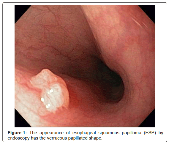
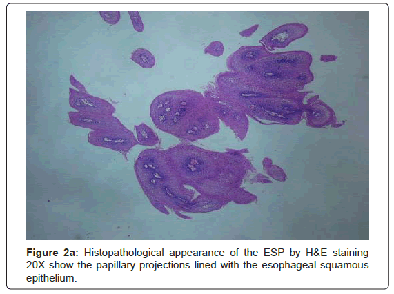
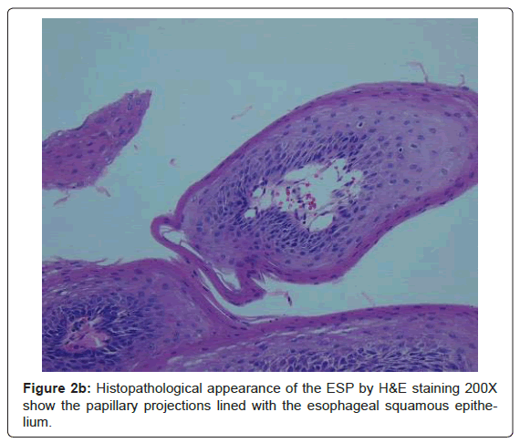
During the upper endoscopy the patient’s esophagus was intubated without difficulty. Tissue surrounding her vocal cords looked erythematous. The esophagus looked normal, without ulcerations, edema, erythema, or furrowing. The lower esophageal sphincter was normal. The stomach and duodenum looked normal. As the scope was withdrawn a small verrucous-looking polyp was noted in the upper esophagus at 20 cm (Figure 1). This was removed by a cold forceps. The size of the polyp was 0.3 x 0.3 cm. There were no immediate complications. The histopathological diagnosis came back as Esophageal Squamous Papilloma (Figure 2a and Figure 2b).
Discussion
ESP is a rare entity that represents a benign tumor in pediatrics and adults [1]. Etiology is not entirely known, it has been proposed that it could be either due to esophagitis from GERD, trauma or Human Papilloma Virus HPV infection [2-4]. Our patient didn’t have any signs of GERD or trauma. There is a likelihood that her EzSP is due to HPV infection although she had no signs of infection else where in her body.
Most cases of ESP are asymptomatic and are found incidentally on EGD [4,5]. Our patient had the feeling that there was something stuck in her throat. This made us think of esophageal eosinophilic disease; however the mid and distal esophageal biopsies were normal.
Recurrence of ESP has not been reported. Multiple papillomatosis rarely progress to squamous cell carcinoma [6]. So as far as we know, there is no need to do surveillance endoscopies to follow up on these lesions.
Conclusion
Endoscopists should be aware of this rare entity and should thoroughly assess the entire esophagus. It will be of interest to follow a cohort of these patients and see the progression of symptoms in the future.
Acknowledgements
We acknowledge the pathology department at the University of Iowa and specifically Dr. Chris Jensen for providing us with the histology images.
References
- Mosca S, Manes G, Monaco R, Bellomo PF, Bottino V, et al. (2001) Squamous papilloma of the esophagus: long-term follow up. J Gastroenterol Hepatol 16: 857-861.
- Afonso LA, Moyses N, Cavalcanti SM (2010) Human papillomavirus detection and p16 methylation pattern in a case of esophageal papilloma. Braz J Med Biol Res 43: 694-696.
- Sandvik AK, Aase S, Kveberg KH, Dalen A, Folvik M, et al. (1996) Papillomatosis of the esophagus. J Clin Gastroenterol 22: 35-37.
- Rebeuh J, Willot S, Bouron-Dal Soglio D, Patey N, Herzog D, et al. (2011) Esophageal squamous papilloma in children. Endoscopy 43: E256.
- Alkhouri N, Wyneski M, Kay M, Wyllie R (2008) Endoscopic appearance of an esophageal squamous papilloma in a pediatric patient. J Pediatr Gastroenterol Nutr 46: 237.
- Attila T, Fu A, Gopinath N, Streutker CJ, Marcon NE (2009) Esophageal papillomatosis complicated by squamous cell carcinoma. Can J Gastroenterol 23: 415-419.
Relevant Topics
- Constipation
- Digestive Enzymes
- Endoscopy
- Epigastric Pain
- Gall Bladder
- Gastric Cancer
- Gastrointestinal Bleeding
- Gastrointestinal Hormones
- Gastrointestinal Infections
- Gastrointestinal Inflammation
- Gastrointestinal Pathology
- Gastrointestinal Pharmacology
- Gastrointestinal Radiology
- Gastrointestinal Surgery
- Gastrointestinal Tuberculosis
- GIST Sarcoma
- Intestinal Blockage
- Pancreas
- Salivary Glands
- Stomach Bloating
- Stomach Cramps
- Stomach Disorders
- Stomach Ulcer
Recommended Journals
Article Tools
Article Usage
- Total views: 16464
- [From(publication date):
July-2012 - Apr 04, 2025] - Breakdown by view type
- HTML page views : 11836
- PDF downloads : 4628


