Review Article Open Access
Development of Anti-Toxins Antibodies for Biodefense
Thibaut Pelat1, Arnaud Avril1, Siham Chahboun1, Jacques Mathieu2 and Philippe Thullier1*
1Unité de biotechnologies des anticorps et des toxines, Département de biologie des agents transmissibles, Institut de recherche biomédicale des armées (IRBA-SSA), 24 avenue des Maquis du Grésivaudan, 38702 La Tronche, France
2Département de biologie des agents transmissibles, Institut de recherche biomédicale des armées (IRBA-CRSSA), 24 avenue des Maquis du Grésivaudan, 38702 La Tronche, France
- *Corresponding Author:
- Philippe Thullier
Unité de biotechnologies des anticorps et des toxines
Département de biologie des agents transmissibles
Institut de recherche biomédicale des armées (IRBA-CRSSA)
24 avenue des Maquis du Grésivaudan, 38702 La Tronche, France
E-mail: pthullier@yahoo.com
Received Date: September 15, 2011; Accepted Date: November 15, 2011; Published Date: November 18, 2011
Citation: Pelat T, Avril A, Chahboun S, Mathieu J, Thullier P (2011) Development of Anti-Toxins Antibodies for Biodefense. J Bioterr Biodef S2:005 doi: 10.4172/2157-2526.S2-005
Copyright: © 2011 Pelat T, et al. This is an open-access article distributed under the terms of the Creative Commons Attribution License, which permits unrestricted use, distribution, and reproduction in any medium, provided the original author and source are credited.
Visit for more related articles at Journal of Bioterrorism & Biodefense
Abstract
Antibodies (Abs) may significantly improve the outcome of diseases caused by toxins of military interest, in particular ricin and botulism toxins. The efficacy of Abs to neutralize ricin was demonstrated in vivo utilizing Abs of animal origin but recombinant antibodies (rAbs), which would be better tolerated, are preferred for clinical use. Animal Abs are utilized at present as medical countermeasure to neutralize botulism toxins, but they show limitations and rAbs are also preferred for this clinical use. Of note, anthrax is an infective disease depending on toxins for its pathogenicity, and it was demonstrated that anti-anthrax toxins Abs could be of benefit when used as adjunct to antibiotics. We used an original strategy, starting from libraries derived from lymphocytes of non-human primates (NHP) immunized with the toxin of interest, to isolate Abs directed against all these toxins. The libraries are screened by the phage-display technology to isolate the best candidates, which are then tested for their toxin neutralization properties. Abs neutralizing the anthrax lethal toxin (35PA83, 2LF) and ricin (43RCA) have been isolated and at present, Abs neutralizing the botulinum toxins (BoNT) A, B and E are being isolated.
Keywords
Antibodies; Anthrax toxins; Ricin; Botulinum toxins; Humanization.
Abbreviations
scFv: Single chain Fv; Fab: Fragment antigenbinding; Ig: Immunoglobulin; PA: Protective antigen; LF: Lethal factor; LT: Lethal toxin.
Introduction
Antibodies of animal origin have been utilized to demonstrate the efficacy of Abs to neutralize toxins, but their use in clinics is limited by their poor tolerance [1], as is the case for the treatment of botulism. The functional equivalents of these animals Abs should be developed in their recombinant form, a class of therapeutic molecules which is presently growing at a rapid pace. The good tolerance of rAbs is the result of their nature, which has become increasingly human over the three successive generations of rAbs - chimerized Abs, humanized Abs and human Abs - that have been introduced in clinical use [2]. The majority of these rAbs are focused against oncological and autoimmune diseases, but recent clinical trials showed an increasing interest in the field of toxin inhibitors and anti-infective drugs [3]. Defense against deliberately caused biological attack (or biodefense) may benefit from the emergence of this new therapeutic class, since Abs have proved to be protective in models of the six diseases often considered most at risk of being caused intentionally [4-5]: ricin poisoning [6], botulinum neurotoxins (BoNTs) [7], anthrax [8], plague [9], tularemia [10] and smallpox [11]. Ricin is a lethal toxin that inhibits protein synthesis. It is easily extracted from an ubiquitously grown plant, Ricinus communis, and thus readily available for use as a bioweapon [12]. BoNTs are regarded as the most toxic substances on Earth, causing naturally-occurring food poisoning, and reaching LD50 values equal to 1 ng/kg for the intravenous and subcutaneous routes or 3 ng/kg for the pulmonary route [13]. Bacillus anthracis, the agent responsible of anthrax, secretes a toxin on which it relies for pathogenesis, and Abs targeting this toxin improve the outcome of the disease.
Obtaining therapeutic antibodies: a review of existing strategies, from polyclonal antibodies to the third generation of recombinant antibodies
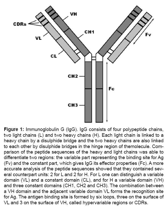
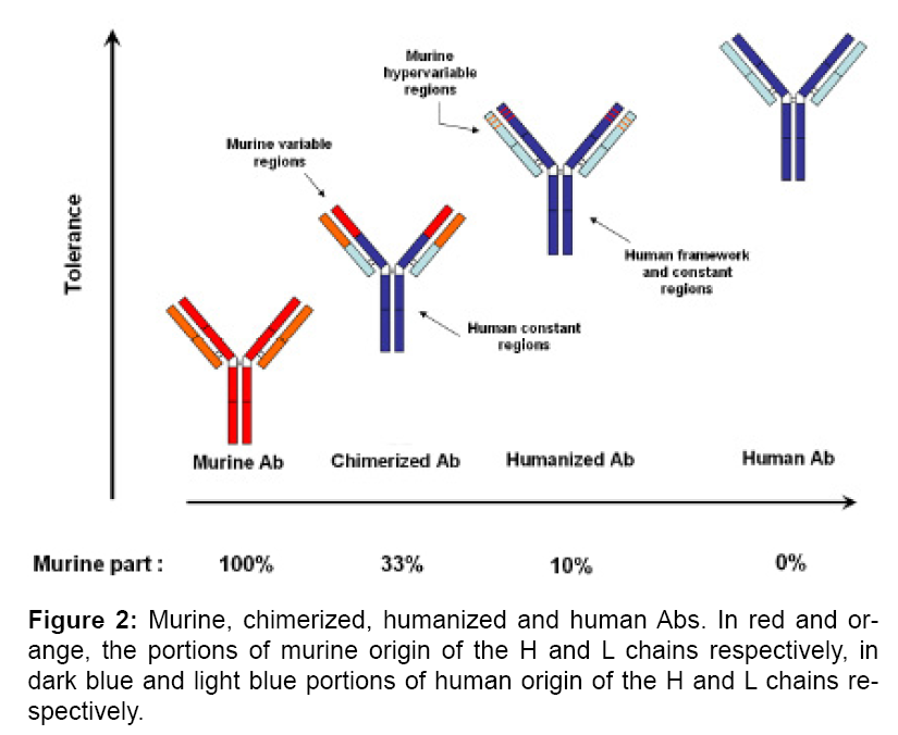
The sera obtained after immunization of animals were used in medicine from the late 18th century. This use was recognized by the first Nobel Prize for Medicine, which was awarded to a German military doctor, Emil von Behring, for developing a serum therapy against diphtheria and tetanus toxins. The benefit of these sera was, however, greatly limited by major intolerance phenomena since they consist of proteins that the human body recognizes as foreign, and which trigger deleterious immune responses, aimed at their elimination. In the mid-20th century, cell biology allowed the isolation of Abs called monoclonal antibodies (mAbs) because each one is secreted identically by one clone of cells, representing in this respect the opposite of polyclonal sera. Hybridoma technology is the technical support for isolating such mAbs, and it earned Köhler and Milstein the Nobel Prize for Medicine in 1984, which was shared with Jerne. MAbs, which are usually immunoglobulins G (IgG, Figure 1), afford advantages in terms of reproducibility and are major research tools. However, these products have hardly contributed anything to medicine due to their animal origin (mostly murine), causing similar intolerance problems as seen with the use of polyclonal sera. At the end of the 20th century, molecular biology techniques made it possible to chimerize and then humanize these murine mAbs, that is, to obtain Abs derived from the previous ones but whose regions of animal origin have been reduced to improve their tolerance (Figure 2). The Abs obtained by these molecular techniques were named recombinant Abs (rAbs), reflecting the fact that DNA from different sources has been combined into a single molecule. The regions of animal origin that have been preserved are those that interact closely with the antigen, i.e. the variable regions in the case of chimerized Abs, or the smaller hypervariable regions in the case of humanized Abs. At the end of the 20th century, two different technological advances made it possible to isolate rAbs independently of animal origin, paving the way for the third generation of rAbs, the fully human Abs. The first of these technological advances was the replacement of the murine chromosomal locus encoding the Abs with its human counterpart, to obtain so-called humanized mice [14,15]. The second was to obtain several very large-sized sets of Abs fragments (scFv or Fab, see Figure 3) called "naïve" libraries (1010 or 1012 clones) [16,17], essentially recapitulating the primary immune response depending on IgMs and directed against all antigens. These libraries can be screened through phage-display technology. However, naïve libraries and humanized mice are very expensive as they were generally developed by private companies (Morphosys in Germany, CAT in Great Britain and Dyax in the United States for naïve libraries; Medarex in the United States and Kirin in Japan for humanized mice), none of which are French and we have sought to become independent to develop rAbs for biodefense.
Figure 1: Immunoglobulin G (IgG). IgG consists of four polypeptide chains, two light chains (L) and two heavy chains (H). Each light chain is linked to a heavy chain by a disulphide bridge and the two heavy chains are also linked to each other by disulphide bridges in the hinge region of themolecule. Comparison of the peptide sequences of the heavy and light chains was able to differentiate two regions: the variable part representing the binding site for Ag (Fv) and the constant part, which gives IgG its effector properties (Fc). A more accurate analysis of the peptide sequences showed that they contained several counterpart units: 2 for L and 2 for H. For L one can distinguish a variable domain (VL) and a constant domain (CL), and for H a variable domain (VH) and three constant domains (CH1, CH2 and CH3). The combination between a VH domain and the adjacent variable domain VL forms the recognition site for Ag. The antigen binding site is formed by six loops, three on the surface of VL and 3 on the surface of VH, called hypervariable regions or CDRs.
Figure 3: Ab fragments smaller in size than a whole Ab can be produced efficiently in E. coli and thus they prove to be better suited to laboratory studies. Stable Fab fragments (50 kDa as against 150 kDa for the whole Ab) have been developed, both chains are kept in the form of heterodimers by the interactions between the variable domains of the heavy and light chains (VH and VL), but above all, by disulphide bridges between the constant domains (CH1 and CL). Smaller fragments called ScFv (25 kDa), consisting of only one VH and one VL linked by a peptide bond (or “linker”), were also developed.
An alternative to naïve libraries is the production of immune libraries deriving from human subjects immunized against the target pathogen. Unlike naïve libraries, one immune library must be built for each of these agents but immune libraries are smaller in size (about 108 clones) than naïve libraries. The advantage of these immune libraries is that they recapitulate the secondary immune response, depending on IgG and thus use the affinity maturation process allowed by the immune system. This is in opposition to naïve libraries, whose Abs often have to be improved by in vitro mutation techniques. It is important to note that the phage-display technology makes it possible to only isolate antibody fragments (scFv or Fab, Figure 3), corresponding to the variable regions [18]. The affinity and neutralizing properties of these Ab fragments can be assessed in vitro. To be tested in vivo, these Ab fragments have to be expressed in fusion with Ab constant regions to obtain whole immunoglobulins G (IgGs), which have a sufficient halflife for these tests.
Obtaining therapeutic antibodies for the treatment of toxininduced diseases belonging to the biological risk by our unit: use of immune libraries derived from NHP
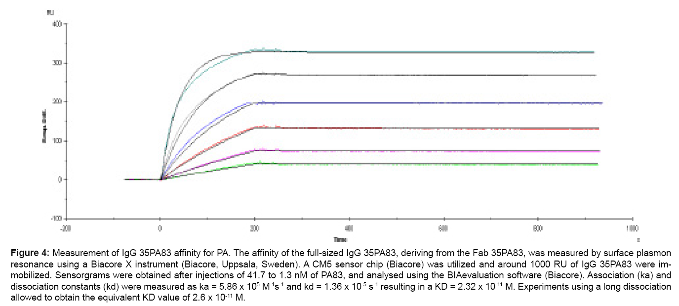
In general, but especially in the case of biodefense and bioterrorism, construction and use of immune libraries is limited by the lack of immunized human subjects, who would be donors of lymphocytes to be used for the construction of these libraries. Macaques (Macaca fascicularis) are primates close to humans and their Abs can be expected to also be close to human Abs [19]. The macaque locus encoding Abs is greater than that of humanized mice, and thus allows for greater diversity and better affinity [20]. In the absence of immunized human donors of lymphocytes and for ethical, theoretical and practical reasons, it is thus advisable to use macaques for the construction of immune libraries. With this approach, we first obtained a Fab fragment directed against tetanus toxin, regarded as a model toxin [21]. Then, using antigens of military interest, we obtained the Fab 35PA83 directed against the protective antigen (PA), one of the two sub-units of the anthrax lethal toxin (LT). Fab 35PA83 presented a high affinity (3.4 nM) and effectively neutralized LT in the standardized neutralization test (IC50 = 5.6 nM) [22]. It was showed that Fab 35PA83 inhibits the interaction of PA with the second toxin sub-unit, blocking the formation of the toxin itself. The affinity of Fab 35PA83 was improved in vitro [23], then the Fab was expressed as a full-sized IgG whose affinity for the protective antigen (PA) was measured as 0.02 nM (Figure 4). Raxibacumab®, a recombinant IgG developed for therapeutic purposes against anthrax, has the same target than IgG 35PA83 and is currently stockpiled under the Bioshield program in the US. It bound its antigen with a 2.78 nM affinity [24]. Anthim® and Thravixa® , other comparable recombinant antibodies also under clinical development, showed affinities of respectively 0.33 nM [25] and 0.08 nM [26] for PA, so it appears that IgG 35PA83 presents the best affinity among those antibodies. IgG 35PA83 neutralization capacity was first tested in a model of anthrax where the A/J strain of mice was utilized. A/J mice lack complement so that the Sterne anthrax strain, which lacks a capsule and is generally regarded as vaccinal, is lethal in these mice. The validity of this model, usable in a level 2 safety laboratory only, is well accepted [27]. In this model, IgG 35PA83 demonstrated its ability to increase the therapeutic window and shorten the duration of antibiotherapy of anthrax.
Figure 4: Measurement of IgG 35PA83 affinity for PA. The affinity of the full-sized IgG 35PA83, deriving from the Fab 35PA83, was measured by surface plasmon resonance using a Biacore X instrument (Biacore, Uppsala, Sweden). A CM5 sensor chip (Biacore) was utilized and around 1000 RU of IgG 35PA83 were immobilized. Sensorgrams were obtained after injections of 41.7 to 1.3 nM of PA83, and analysed using the BIAevaluation software (Biacore). Association (ka) and dissociation constants (kd) were measured as ka = 5.86 x 10⁵ M-1s-1 and kd = 1.36 x 10⁻⁵ s-1 resulting in a KD = 2.32 x 10⁻¹¹ M. Experiments using a long dissociation allowed to obtain the equivalent KD value of 2.6 x 10⁻¹¹ M.
In effect, it was showed that when antibioprophylaxis using doxycycline was performed for only 7 days after exposure to anthrax, the mice died soon after this treatment was stopped. However, when 35PA83 IgG (2 mg/kg) was added to the antibioprophylaxis on the 7th day, all mice survived. Also, when a treatment exclusively consisting of ciprofloxacin was started 12 hours after exposure to anthrax, no animal survived. When 35PA83 IgG was added to ciprofloxacin, 80% of the animals survived, corresponding to an increased therapeutic window. IgG 35PA83 was also tested in a second anthrax model utilizing New Zealand rabbits, infected with a lethal strain of anthrax isolated in a fatal human case in France [28], thus requesting a level 3 safety laboratory. In this model, which is standard for testing antibody efficacy against anthrax, a single dose of 2.5 mg/kg of IgG 35PA83 coadministered with the spores protected all infected animals. In this rabbit model but using another lethal strain of anthrax (Ames), 40mg/ kg of Raxibacumab®, or doses of 4 mg/kg of Anthima® or of Thravixa® [29], were requested for an equivalent result thus also reflecting an apparently greater efficacy of IgG 35PA83. Isolation of the following Fab fragments was marked by difficulties related to the instability of the libraries, and we moved to produce Ab fragments in scFv format, which is smaller than the Fab format (Figure 3). This has enabled us to isolate an scFv, 2LF, neutralizing the LT but directed against its second sub-unit, the lethal factor (LF), in accordance with the anthrax experts recommendations for future treatments [30,31]. 2LF development aims at preventing any risk of LT escaping anti-PA antibodies, by natural or induced mutation. Synergies with the anti-PA will be tested, given that such synergies between anti-PA and anti-LF have already been observed [32]. ScFv 2LF has a high affinity to LF (KD = 1.02 nM) and is highly neutralizing in the standardized in vitro assay as well as in vivo assay [31]. Its epitope was localized in the domain I of LF and thus 2LF inhibits the formation of the LF-PA complex, i.e. the LT formation. 2LF was the first recombinant fragment of Ab having this activity and it has just been expressed in the form of IgG for in vivo testing, similarly as for the IgG derived from Fab 35PA83. In addition, we isolated an scFv, 43RCA, of very high affinity to ricin toxin (KD = 40 pM), which is one of the best affinities ever published without in vitro maturation. ScFv 43RCA neutralized ricin in vitroand in vivo [33]. This scFv was expressed as IgG and tested in mice intoxicated by instillation of ricin. IgG 43RCA, also administered by instillation, remained completely effective 6 hours after this intoxication (Pr K.- M. Chu-Wang, University of Medicine, New York, USA, personal communication). Recently, we received European funding for a project, AntibotABE (www.antibotabe.com), aimed at isolating scFvs against the heavy chain (Hc region) and the light chain of the three botulinum toxin serotypes highly pathogenic for humans, BoNT/A, B and E. The same strategy as previously will be tilized. The AntiBotABE consortium consists of nine partners, academic teams and private companies from four different countries, and aims at expressing these scFvs as recombinant IgGs, of potential value for biodefense in four years. Until now, NHP immune libraries directed against Hc fragments of BoNT/A, B, E and the light chain of BoNT/A have been panned, and neutralizing scFvs are sought among the most affine candidates. Macaques are still undergoing immunization with the light chain of BoNT/B.
Engineering of NHP antibodies: presentation and clinical value of germline humanization, towards the fourth generation of recombinant antibodies?
Our strategy is very efficient to isolate neutralizing Abs, which however are not human but derive from NHPs. We have shown that there are differences between human and NHP Abs, so that NHP Abs have to be humanized [34,35]. For this humanization, we use human germline sequences as templates because they are part of the human immunological self and should thus be optimally tolerated. This is in opposition to human IgGs, which are mutated during the affinity maturation process and do not represent molecules that are always well tolerated, as shown recently by adalimumab [36,37]. Our approach is thus to "germline-humanize" i.e. to mutate Ab variable regions to make them closer to human germline sequences. For instance the framework regions of parental 35PA83, which were 87.6% identical to their closer human germline sequences (retrieved using IMGT tools [38]), were germline-humanized, resulting in a degree of identity increased to 97.8% [39]. The germline-humanized version of 35PA83 is thus very similar to human germline sequences and likely to be very well tolerated, while retaining its affinity and its neutralization properties. An identical process was applied to 2LF and 43RCA with similar promising results. On the particular case of 43RCA, it was verified by Antitope (Cambridge, United Kingdom) that germlinehumanization had suppressed all T epitopes, and the immunogenicity of the germlinized 43RCA in Humans was therefore predicted to be alsosuppressed. Multiple injections of germline-humanized Abs should not raise an immune response so that these Abs could be used for prophylaxis. In fact, germline-humanized Abs could represent the fourth generation of rAbs.
Conclusion
Antibodies neutralizing ricin, anthrax toxins and more recently botulinum 203 toxins have been isolated by a strategy consisting of building and screening phage-displayed libraries derived from immunized macaques, then of humanizing the best candidates. The excellent tolerance of our antibodies should allow them to be used not only for therapy, but also for prophylaxis.
Acknowledgements
This review was made possible by Direction Générale de l'Armement funding, under n° 09co302-1.
References
- Black RE, Gunn RA (1980) Hypersensitivity reactions associated with botulinal antitoxin. Am J Med 69: 567-570.
- Bellet D, Dangles-Marie V (2005) [Humanized antibodies as therapeutics] Med Sci (Paris) 21: 1054-1062.
- Pai JC, Sutherland JN, Maynard JA (2009) Progress towards recombinant antiinfective antibodies. Recent Pat Antiinfect Drug Discov 4: 1-17.
- Thullier P, Pelat T, Vidal D (2009) [Recombinant antibodies against bioweapons]. Med Sci (Paris) 25: 1145-1148.
- Froude JW, Stiles B, Pelat T, Thullier P (2011) Antibodies for biodefense. mAbs 3: 517-527
- Pratt TS, Pincus SH, Hale ML, Moreira AL, Roy CJ, et al. (2007) Oropharyngeal aspiration of ricin as a lung challenge model for evaluation of the therapeutic index of antibodies against ricin A-chain for post-exposure treatment. Exp Lung Res 33: 459-481.
- Rodriguez GC, Geren IN, Lou J, Conrad F, Forsyth C, et al. (2011) Neutralizing human monoclonal antibodies binding multiple serotypes of botulinum neurotoxin. Protein Eng Des Sel 24: 321-331.
- Subramanian GM, Cronin PW, Poley G, Weinstein A, Stoughton SM, et al. (2005) A phase 1 study of PAmAb, a fully human monoclonal antibody against Bacillus anthracis protective antigen, in healthy volunteers. Clin Infect Dis 41:12-20.
- Xiao X, Zhu Z, Dankmeyer JL, Wormald MM, Fast RL, et al. (2010) Human antiplague monoclonal antibodies protect mice from Yersinia pestis in a bubonic plague model. Plos one 5: e13047.
- Lu Z, Roche MI, Hui JH, Unal B, Felgner PL, et al. (2007) Generation and characterization of hybridoma antibodies for immunotherapy of tularemia.Immunol Lett 112: 92-103.
- McCausland MM, Benhnia MR, Crickard L, Laudenslager J, Granger SW, et al. (2010) Combination therapy of vaccinia virus infection with human anti-H3 and anti-B5 monoclonal antibodies in a small animal model. Antivir Ther 15:661-675.
- Audi J, Belson M, Patel M, Schier J, Osterloh J (2005) Ricin poisoning: a comprehensive review. Jama 294: 2342-2351.
- Gill DM (1982) Bacterial toxins: a table of lethal amounts. Microbiol Rev 46: 86-94.
- Bruggemann M, Neuberger MS (1996) Strategies for expressing human antibody repertoires in transgenic mice. Immunol Today 17: 391-397.
- Tomizuka K, Shinohara T, Yoshida H, Uejima H, Ohguma A, et al. (2000) Double trans chromosomic mice: maintenance of two individual human chromosome fragments containing Ig heavy and kappa loci and expression of fully human antibodies. Proc Natl Acad Sci U S A 97: 722-727.
- Vaughan TJ, Williams AJ, Pritchard K, Osbourn JK, Pope AR, et al. (1996) Human antibodies with sub-nanomolar affinities isolated from a large nonimmunized phage display library. Nat Biotechnol 14: 309-314.
- Little M, Welschof M, Braunagel M, Hermes I, Christ C, et al. (1999) Generation of a large complex antibody library from multiple donors. J Immunol Methods 231: 3-9.
- Barbas CF (2001) Phage display : a laboratory manual. Cold Spring Harbor NY Cold Spring Harbor Laboratory Press.
- Andris-Widhopf J, Steinberger P, Fuller R, Rader C, Barbas CF (2001) Protocol 9.36: Librairies of nonhuman primate anibody fragments. In Phage display : a laboratory manual. Edited by Barbas CF. Cold Spring Harbor, NY: Cold Spring Harbor Laboratory Press.
- Pelat T, Hust M, Thullier P (2009) Obtention and engineering of non-human primate (NHP) antibodies for therapeutics. Mini Rev Med Chem 9: 1633-1638.
- Chassagne S, Laffly E, Drouet E, Herodin F, Lefranc MP, et al. (2004) A highaffinity macaque antibody Fab with human-like framework regions obtained from a small phage display immune library. Mol Immunol 41: 539-546.
- Laffly E, Danjou L, Condemine F, Vidal D, Drouet E, et al. (2005) Selection of a macaque Fab with framework regions like those in humans, high affinity, and ability to neutralize the protective antigen (PA) of Bacillus anthracis by binding to the segment of PA between residues 686 and 694. Antimicrob Agents Chemother 49: 3414-3420.
- Laffly E, Pelat T, Cedrone F, Blesa S, Bedouelle H, et al. (2008) Improvement of an antibody neutralizing the anthrax toxin by simultaneous mutagenesis of its six hypervariable loops. J Mol Biol 378:1094-1103.
- Mazumdar S (2009) Raxibacumab. Mabs 1: 531-538.
- Mohamed N, Clagett M, Li J, Jones S, Pincus S, et al. (2005) A high-affinity monoclonal antibody to anthrax protective antigen passively protects rabbits before and after aerosolized Bacillus anthracis spore challenge. Infect Immun 73: 795-802.
- Sawada-Hirai R, Jiang I, Wang F, Sun SM, Nedellec R, et al. (2004) Human anti-anthrax protective antigen neutralizing monoclonal antibodies derivedfrom donors vaccinated with anthrax vaccine adsorbed. J Immune Based Ther Vaccines 2: 5.
- Welkos SL, Keener TJ, Gibbs PH (1986) Differences in susceptibility of inbred mice to Bacillus anthracis. Infect Immun 51: 795-800.
- Berthier M, Fauchere JL, Perrin J, Grignon B, Oriot D (1996) Fulminant meningitis due to Bacillus anthracis in 11-year-old girl during Ramadan. Lancet 347: 828.
- Froude JW, Stiles B, Pelat T, Thullier P (2011) Antibodies for biodefense. Mabs 3: 517-527.
- Baillie LWJ (2006) Past, imminent and future human medical countermeasures for anthrax. J Appl Microbiol 101: 594-606.
- Pelat T, Hust M, Laffly E, Condemine F, Bottex C, et al. (2007) High-affinity, human antibody-like antibody fragment (single-chain variable fragment) neutralizing the lethal factor (LF) of Bacillus anthracis by inhibiting protective antigen-LF complex formation. Antimicrob Agents Chemother 51: 2758-2764.
- Brossier F, Levy M, Landier A, Lafaye P, Mock M (2004) Functional analysis of Bacillus anthracis protective antigen by using neutralizing monoclonal antibodies. Infect Immun 72: 6313-6317.
- Pelat T, Hust M, Hale M, Lefranc MP, Dubel S, et al. (2009) Isolation of a human-like antibody fragment (scFv) that neutralizes ricin biological activity.BMC Biotechnol 9: 60.
- Thullier P, Huish O, Pelat T, Martin AC (2010) The humanness of macaque antibody sequences. J Mol Biol 396: 1439-1450.
- Thullier P, Chahboun S, Pelat T (2010) A comparison of human and macaque (Macaca mulatta) immunoglobulin germline V regions and its implications for antibody engineering. Mabs 2: 528-538.
- Bartelds GM, de Groot E, Nurmohamed MT, Hart MH, van Eede PH, et al. (2010) Surprising negative association between IgG1 allotype disparity and anti-adalimumab formation: a cohort study. Arthritis Res Ther 12: R221.
- Van Schouwenburg PA, Bartelds GM, Hart MH, Aarden L, Wolbink GJ, et al. (2010) A novel method for the detection of antibodies to adalimumab in the presence of drug reveals "hidden" immunogenicity in rheumatoid arthritis patients.J Immunol Methods 362: 82-88.
- Giudicelli V, Chaume D, Lefranc MP (2004) IMGT/V-QUEST, an integrated software program for immunoglobulin and T cell receptor V-J and V-D-J rearrangement analysis. Nucleic Acids Res 32: W435-440.
- Pelat T, Bedouelle H, Rees AR, Crennell SJ, Lefranc MP, et al. (2008) Germline humanization of a non-human primate antibody that neutralizes the anthrax toxin, by in vitro and in silico engineering. J Mol Biol 384: 1400-1407.
Relevant Topics
- Anthrax Bioterrorism
- Bio surveilliance
- Biodefense
- Biohazards
- Biological Preparedness
- Biological Warfare
- Biological weapons
- Biorisk
- Bioterrorism
- Bioterrorism Agents
- Biothreat Agents
- Disease surveillance
- Emerging infectious disease
- Epidemiology of Breast Cancer
- Information Security
- Mass Prophylaxis
- Nuclear Terrorism
- Probabilistic risk assessment
- United States biological defense program
- Vaccines
Recommended Journals
Article Tools
Article Usage
- Total views: 14560
- [From(publication date):
specialissue-2014 - Apr 05, 2025] - Breakdown by view type
- HTML page views : 9940
- PDF downloads : 4620


