Complex Concha Bullosa
Received: 03-Feb-2012 / Accepted Date: 25-Feb-2012 / Published Date: 01-Mar-2012 DOI: 10.4172/2161-119X.1000109
250880Introduction
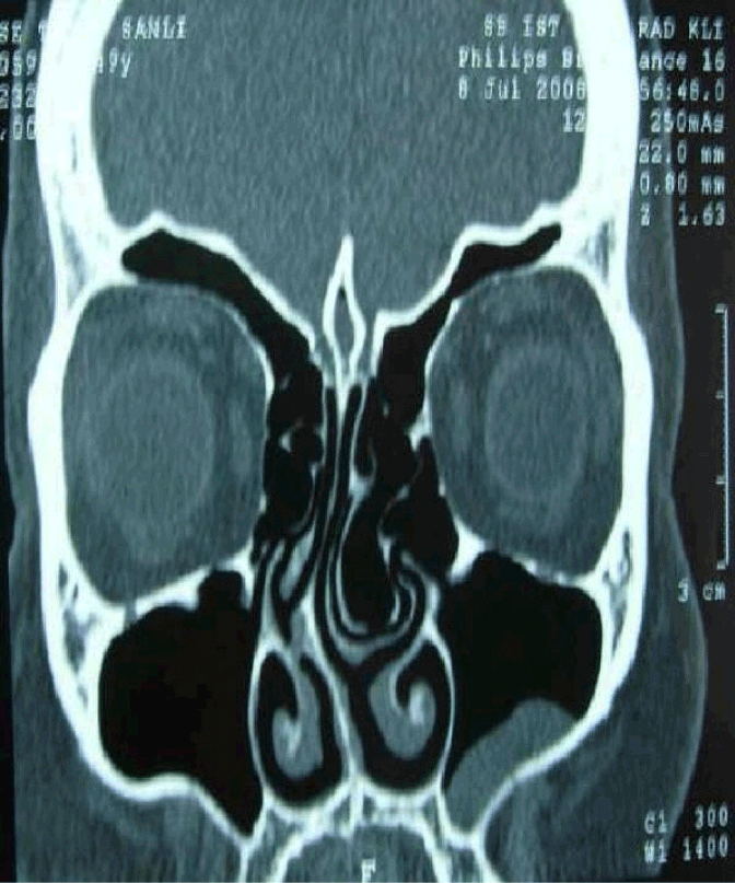
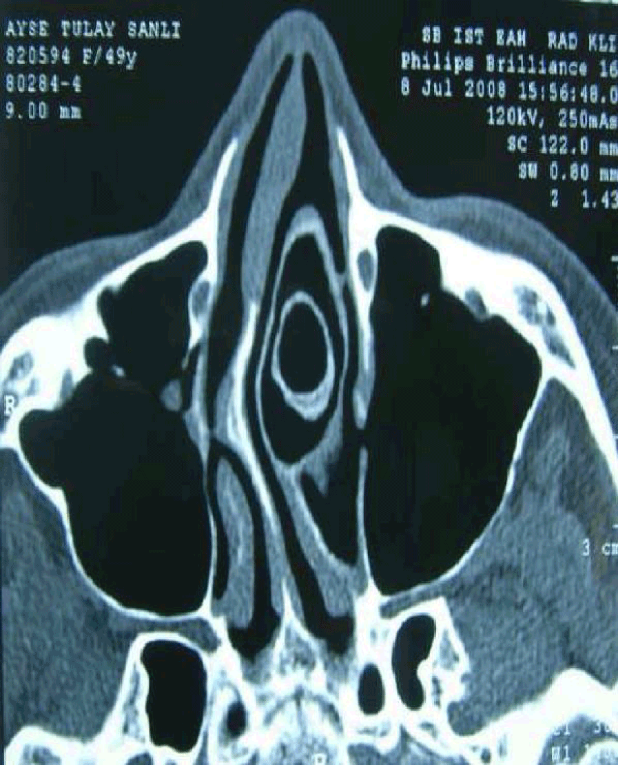
As endoscopic sinus surgery has been used widely in sinus pathologies and advancements in computerized tomography technologies, the importance of the lateral nasal wall anatomy and its variations has been emphasized more commonly. In evaluation of endoscopic and radiological anatomy of paranasal sinuses attention must be paid to these variations. Concha bullosa which is an anatomical variation of paranasal region, is pneumatization of middle conchal bone. Concha bullosa can be uni- or bilateral and if it is unilateral, it is accompanied usually by a contralateral septal deviation. Ethmoid bulla is an anterior ethmoidal cell. Its size, shape and drainage may show differences among people. In this case we present a big ethmoid bulla invaginating into a giant concha bullosa.
Case Presention
A 49 year-old female patient was admitted to our clinic with complaints of nasal obstruction, facial pain and snoring. During anterior rinoscopy right sided septal deviation and left sided nasal mass with a smooth mucosal surface was noted. The mass was compatible with concha bullosa and almost completely obstructing the nasal cavity. We failed to perform nasal endoscopy to the left side. Paranasal sinus tomography in axial and coronal sections demonstrated a left sided big ethmoid bulla invaginating into a giant concha bullosa (Figure 1and 2). An endoscopic concha bullosa and ethmoid bulla resection with a septoplasty was performed. Patient’s complaints disappeared after surgery.
Discussion
With advances of endoscopic techniques, progresses have been increased in paranasal sinus surgery. On the other hand this region has many anatomical variations which increase the risk of complications of endoscopic surgery. In addition to endoscopic examination paranasal sinus tomography in axial and coronal sections are very important for demonstrating both the pathology and related anatomic regions. The most common anatomical variation in the region of osteomeatal complex is concha bullosa. It’s prevalence change from 13.2% to 72.2% in different articles [1-7] and between 33.8% and 72.6% in symptomatic cases respectively [4].
Concha bullosa has three variations according to its location and shape of pneumatization [4].
1. Lamellar form: pneumatization is limited to its vertical lamella (45- 46%).
2. Bulbous form: pneumatization is in its inferior part (21-31%).
3. Big form: a massive pneumatization is in both vertical lamella and inferior part (16-34%).
Concha bullosa is generally small and asymptomatic. But if it reaches bigger sizes it can disrupt sinus drainage and nasal air flow. Concha bullosa has been related to sinusitis in proportion to its size, however there are some studies which defend the opposite [8,9]. Etiology of concha bullosa formation is not exactly. According to Stammberger and Braun [10], pneumatization of middle concha can result from middle meatus, frontal recess, suprabulbar and retrobulbar recesses and less likely ethmoid infindibulum and agger nasi region.
There are two theories for situations in which septal deviation and concha bullosa coexist [11]. According to first one, the cause of concha bullosa formation is septal deviation. Cavity in wide side will be filled by concha bullosa. Second theory suggests that nasal septum deviates because of concha bullosa. There is no absolute evidence for both theories. Ethmoid bone is with no doubt one of the most complicated anatomic structures. Its cells are named anterior and posterior cells in respect of drainage regions. However both anterior and also posterior cells show too many variations. Ethmoid bulla is located in anterior region and its classification is controversial. Although it is defined as a sinus cell, due to lack of its posterior bony wall it could also be considered as a bony lamella as suggested by Wright and Bolger. Scribano et al. [12] showed a 5.4% variation of big ethmoid bulla in their study. We could not find a similar case in literature review, except a case report which was published by Köybaşı et al. [13]. In this study bilateral concha bullosa and ethmoid bulla within this concha bullosa was presented and defined as complex concha bullosa.
Setliff et al. [14] has classified ethmoid bulla as simple (47%), compound (26%) and complex (27%) considering its relations to ethmoid cells. They did it with the help of intraoperative video records and defined ethmoid bulla as complex, if there is an extra cell inside it. In light of foregoing studies, we considered that it would be appropriate to define our case as complex concha bullosa. In patient’s left nasal cavity there was a big concha bullosa which was big enough to obstruct the nasal passage and inside it an invagination towards ethmoid bulla. Relationship between concha bullosa and sinusitis or septal deviation has been a subject to many studies. Aktas et al. [15] found a statistically significant relationship between concha bullosa and septal deviation.
Uygur et al. [16] claimed that septal deviation does not cause concha bullosa but it increases pneumatization of concha bullosa in concordance with its angle of deviation. Stallman et al. [17] claimed that there is an important togetherness of concha bullosa and septal deviation but there isn’t a cause-effect relationship due to existence of air passage between concha bullosa and septum. Furthermore, they said that this togetherness does depend on neither concha bullosa size nor severeness of deviation.
Although concha bullosa is associated with sinusitis according to its size, there are also studies against this argument. In a study of Yousem et al. [18], not the existence of concha bullosa but its size was held responsible for occurence of sinusitis. Later in following studies sinusitis was not associated with concha bullosa but concha bullosa was emphasized as a possible cause of sinusitis due to forming mucosal contact and obstruction in osteomeatal complex region [19].
The patient’s complaints had disappeared after endoscopic removal of concha bullosa, resection of ethmoid bulla, and septoplasty.
Conclusion
Anatomical variations of paranasal sinuses are very common. Before performing an endoscopic surgery, nasal anatomy must be studied thoroughly for both pathology and anatomical variations. This giant complex concha bullosa in which ethmoid bulla invaginating is interesting and important as it is defined second time in literature.
References
- Zinreich SJ, Kennedy DW, Rosenbaum AE, Gayler BW, Kumar AJ, et al. (1987) Paranasal sinuses: CT imaging requirements for endoscopic surgery. Radiology 163: 769-775.
- Zinreich SJ, Mattox DE, Kennedy DW, Chisholm HL, Diffley DM, et al. (1988) Concha bullosa: CT evaluation. J Comput Assist Tomogr 12: 778-784.
- Clark ST, Babin RW, Salazar J (1989) The incidence of concha bullosa and its relationship to chronic sinonasal disease. Am J Rhinol 3: 11-12.
- Bolger WE, Butzin CA, Parsons DS (1991) Paranasal sinus bony anatomic variations and mucosal abnormalities: CT analysis for endoscopic sinus surgery. Laryngoscope 101: 56-64.
- Calhoun KH, Waggenspack GA, Simpson CB, Hokanson JA, Bailey BJ (1991) CT evaluation of the paranasal sinuses in symptomatic and asymptomatic populations. Otolaryngol Head Neck Surg 104: 480-483.
- Meloni F, Mini R, Rovasio S, Stomeo F, Teatini GP (1992) Anatomic variations of surgical importance in ethmoid labyrinth and sphenoid sinus. A study of radiological anatomy. Surg Radiol Anat 14: 65-70.
- Cannon CR (1994) Endoscopic management of concha bullosa. Otolaryngol Head Neck Surg 110: 449-454.
- ShechtmanFG, Kraus WM, Schaefer SD (1993) Inflammatory diseases of the sinuses: anatomy. Otolaryngol Clin North Am 26: 509-516.
- Jones NS, Strobl A, Holland I (1997) A study of the CT findings in 100 patients with rhinosinusitis and 100 control. Clin Otolaryngol Allied Sci 22: 47-51.
- Braun H, Stammberger H (2003) Pneumatization of turbinates. Laryngoscope 113: 668-672.
- Stammberger H (1991) Functional endoscopic sinus surgery, the Messerklinger technique. Philadelphia: Decker.
- Wright ED, Bolger WE (2001) The bulla ethmoidalis: lamella or a true cell. J Otolaryngol 30: 162-166.
- Köybasi S, Gürel K, Akyürek F (2005) Kompleks Konka Bülloza. Türk Otolarengololoji Arsivi 43: 229-232.
- Setliff RC 3rd, Catalano PJ, Catalano LA, Francis C (2001) An anatomic classification of the ethmoidal bulla. Otolaryngol Head Neck Surg 125: 598-602.
- Aktas D, Kalcioglu MT, Kutlu R, Özturan O, Öncel S (2003) The relationship between the concha bullosa, nasal septal deviation and sinusitis. Rhinology 41: 103-106.
- Uygur K, Tuz M, Dogru H (2003) The correlation between septal deviation and concha bullosa. Otolaryngol Head Neck Surg 129: 33-36.
- StallmanJS, Lobo JN, Som PM (2004) The incidence of concha bullosa and its relationship to nasal septal deviation and paranasal sinus disease. AJNR Am J Neuroradiol 25:1613-1618.
- Yousem DM, Kennedy DM, Rosenberg S (1991) Osteomeatal complex risk factors for sinusitis: CT evaluation. J Otolaryngol 20: 419-424.
- Scribano E, Ascenti G, Loria G, Cascio F, Gaeta M (1997) The role of osteomeatal unit anatomic variations in inflamatory disease of the maxillary sinuses. Eur J Radiol 24: 172-174.
Citation: Ceylan S, Bora F, Batmaz T, Atalay B (2012) Complex Concha Bullosa. Otolaryngol 2:109. DOI: 10.4172/2161-119X.1000109
Copyright: © 2012 Ceylan S, et al. This is an open-access article distributed under the terms of the Creative Commons Attribution License, which permits unrestricted use, distribution, and reproduction in any medium, provided the original author and source are credited.
Select your language of interest to view the total content in your interested language
Share This Article
Recommended Journals
Open Access Journals
Article Tools
Article Usage
- Total views: 15907
- [From(publication date): 3-2012 - Jul 06, 2025]
- Breakdown by view type
- HTML page views: 11297
- PDF downloads: 4610


