OMICS International organises 3000+ Global Conferenceseries Events every year across USA, Europe & Asia with support from 1000 more scientific Societies and Publishes 700+ Open Access Journals which contains over 50000 eminent personalities, reputed scientists as editorial board members.
Open Access Journals gaining more Readers and Citations
700 Journals and 15,000,000 Readers Each Journal is getting 25,000+ Readers
Clinical Research on Foot & Ankle received 1112 citations as per Google Scholar report
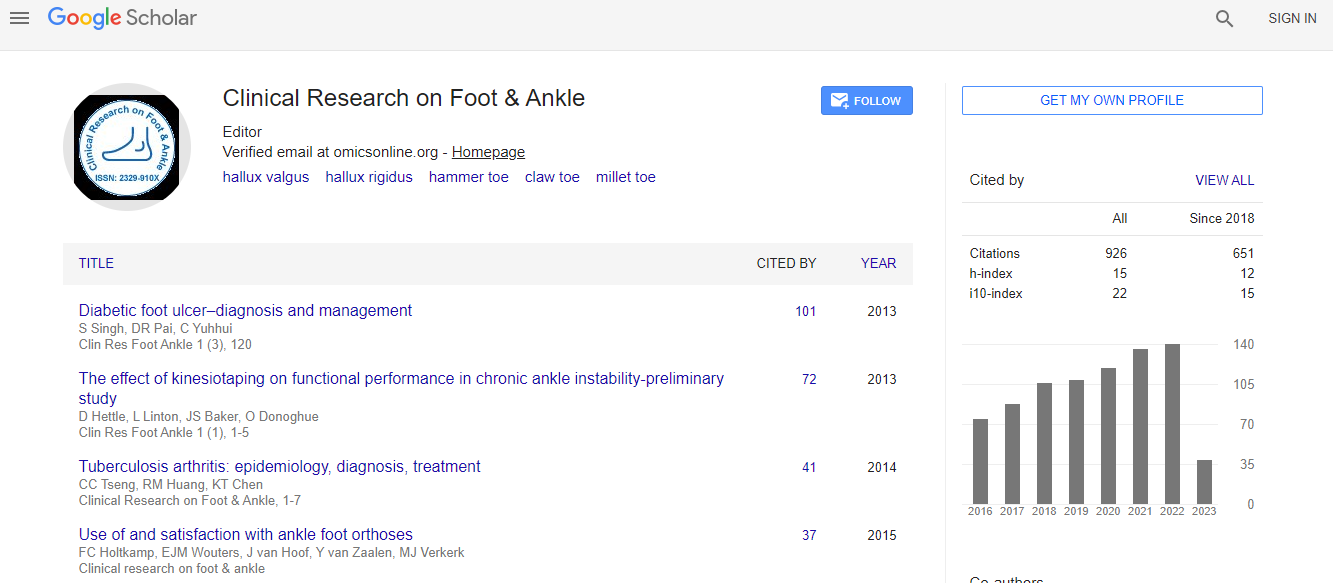

Articles published in Clinical Research on Foot & Ankle have been cited by esteemed scholars and scientists all around the world. Clinical Research on Foot & Ankle has got h-index 15, which means every article in Clinical Research on Foot & Ankle has got 15 average citations.
Following are the list of articles that have cited the articles published in Clinical Research on Foot & Ankle.
| 2024 | 2023 | 2022 | 2021 | 2020 | 2019 | 2018 | 2017 | 2016 | |
|---|---|---|---|---|---|---|---|---|---|
Total published articles |
112 | 88 | 59 | 59 | 18 | 10 | 28 | 31 | 44 |
Research, Review articles and Editorials |
67 | 85 | 38 | 0 | 9 | 9 | 0 | 0 | 0 |
Research communications, Review communications, Editorial communications, Case reports and Commentary |
6 | 7 | 11 | 0 | 2 | 0 | 0 | 0 | 0 |
Conference proceedings |
0 | 0 | 0 | 0 | 0 | 0 | 0 | 0 | 0 |
Citations received as per Google Scholar, other indexing platforms and portals |
76 | 121 | 132 | 137 | 121 | 121 | 105 | 88 | 76 |
| Journal total citations count | 1112 |
| Journal impact factor | 2.34 |
| Journal 5 years impact factor | 3.49 |
| Journal cite score | 3.44 |
| Journal h-index | 15 |
| Journal h-index since 2019 | 12 |
Studying major amputations in a developing country using Amit Jainâs typing and scoring system for diabetic foot complications - time for standardization of diabetic foot practice |
|
| View at Publisher | View at Google Scholar | View at Indexing | |
Antibiotic therapy for diabetic foot infections in a tertiary care hospital in Jakarta, Indonesia |
|
| View at Publisher | View at Google Scholar | View at Indexing | |
Health education programmes to improve foot self-care practices and foot problems among older people with diabetes: a systematic review |
|
| View at Publisher | View at Google Scholar | View at Indexing | |
A retrospective microbiologic study on admitted patients with diabetic foot infection in Razi teaching hospital in Ahvaz during 2015 â2019 |
|
| View at Publisher | View at Google Scholar | View at Indexing | |
LIAISON BETWEEN METHYLENE TETRA HYDRO FOLATE REDUCTASE (MTHFR) ENZYME SINGLE NUCLEOTIDE POLYMORPHISM (C667T) (RS180113) AND DIABETIC SEPTIC FOOT |
|
| View at Publisher | View at Google Scholar | View at Indexing | |
Skrining Aktivitas Antibakteri Bakteriosin dari MinumanCe Hun Tiau |
|
| View at Publisher | View at Google Scholar | View at Indexing | |
Adrenal cortical carcinoma: pathology, genomics, prognosis, imaging features, and mimics with impact on management |
|
| View at Publisher | View at Google Scholar | View at Indexing | |
DIAGNOSTIC OF DIABETIC FOOT ULCER |
|
| View at Publisher | View at Google Scholar | View at Indexing | |
The relationship between knowledge and behavior on the degree of foot injury in type 2 diabetes mellitus patients at Mitra Keluarga Hospital, East Bekasi |
|
| View at Publisher | View at Google Scholar | View at Indexing | |
VITAMIN D DAN DISFUNGSI ENDOTEL PADA KAKI DIABETIK |
|
| View at Publisher | View at Google Scholar | View at Indexing | |
The Burden of Diabetic Foot Disorders on the Patient and the Methods of Treatment |
|
| View at Publisher | View at Google Scholar | View at Indexing | |
The Effect of Modified Diabetes Self-management Education and Support on Self-care and Quality of Life among Patients with Diabetic Foot Ulcers in Rural Area of Indonesia |
|
| View at Publisher | View at Google Scholar | View at Indexing | |
The Burden of Diabetic Foot Disorders on the Patient and the Methods of Treatment |
|
| View at Publisher | View at Google Scholar | View at Indexing | |
Study of Papaya Dressing in Wound Bed Preparation of Diabetic WoundâA Review |
|
| View at Publisher | View at Google Scholar | View at Indexing | |
Plantar temperature and vibration perception in patients with diabetes: A cross-sectional study |
|
| View at Publisher | View at Google Scholar | View at Indexing | |
Unusually delayed manifestation of a hallux osteoid osteoma: A case report Search ScienceDirect Outline Highlights Abstract Keywords 1. Introduction 2. Case presentation 3. Discussion 4. Conclusion Sources of funding Ethical approval Consent Author contribution Registration of research studies Guarantor Provenance and peer review Declaration of Competing Interest References Figures (5) Fig. 1. Anteroposterior and lateral-oblique foot radiographs demonstrating an osteoid⦠Fig. 2. Clinical photograph of the swollen right hallux with erythema Fig. 3. Coronal T1- and fat suppressed T2-weighted (a, b) and axial T1- and fat⦠Fig. 4. Coronal (a) and sagittal (b) fat suppressed T2-weighted magnetic resonance⦠Fig. 5. Consecutive coronal (a, b), axial (c) and sagittal (d) computed tomography⦠Elsevier International Journal of Surgery Case Reports Volume 68, 2020, Pages 8-11 International Journal of Surgery Case Reports Case Report Unusually delayed manifestation of a hallux osteoid osteoma: A case report Author links open overlay panelMeltemÃzdemiraRasime PelinKavakaBegümDemirler ?im?iraEvrimDumanb https://doi.org/10.1016/j.ijscr.2020.02.032Get rights and content Under a Creative Commons licenseopen access Highlights ⢠Osteoid osteoma typically occurs in children or in adolescents. However, it should be noted that this tumor can be detected in older age groups as well. ⢠Although extremely rare, osteoid osteoma may occur in the hallux. ⢠CT is the method of choice in the diagnosis of osteoid osteoma. ⢠Osteoid osteoma should be included in the differential diagnosis list in all patients presenting with foot pain. Abstract Introduction Osteoid osteomas are benign osteoblastic bone tumors mostly seen in patients in the second or third decade of life, and they most frequently involve the femur and tibia. Hallux osteoid osteoma is an extremely rare occurrence with only 14 reported cases to date. Presentation of case A 46-year-old woman with a right hallux pain for the last 18 months was admitted. Her foot radiographs showed a small sclerotic focus on the distal phalanx and degenerative changes in the interphalangeal joint of the hallux. The complaints of the patient were attributed to osteoarthritis involving the interphalangeal joint and non-steroidal anti-inflammatory drugs were administered; however, the patientâs condition did not improve, and hallux osteoid osteoma could not be correctly diagnosed until a CT scan was performed. Discussion We present a case of hallux osteoid osteoma in which the diagnosis was delayed greatly due to the unusuality of the lesion location, the advanced age of the patient, and the uncertainty of the clinical and radiographic appearance. Conclusion Although rare, osteoid osteoma may occur in the hallux. Even if the patient age, pain pattern, and radiographic findings do not exactly meet the classical definitions for osteoid osteoma, this tumor should always be included in the differential diagnosis list in patients presenting with foot pain. |
|
| View at Publisher | View at Google Scholar | View at Indexing | |
Juvenile Osteoid Osteoma of the Distal Phalanx of the Second Digit: A Case Report |
|
| View at Publisher | View at Google Scholar | View at Indexing | |
Juvenile Osteoid Osteoma of the Distal Phalanx of the Second Digit: A Case Report |
|
| View at Publisher | View at Google Scholar | View at Indexing | |
Make the best use of Scientific Research and information from our 700 + peer reviewed, Open Access Journals