|
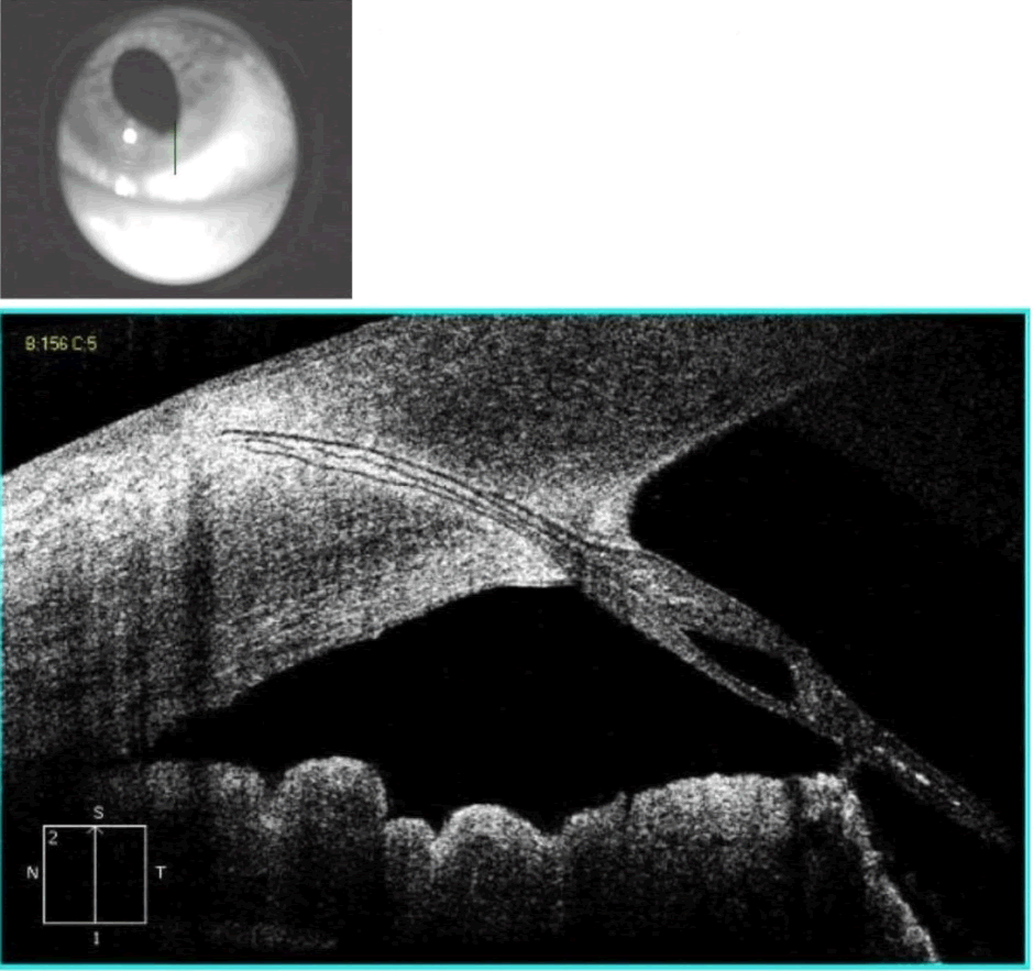
| Figure 1: Cirrus SD-OCT anterior segment vertical scan. Vitreous strand is seen prolapsing through the pupil, attaching to the iris and penetrating into the cornea to the level of the stroma at the incisional wound site. The epithelium is intact and no external vitreous strand is visible. |