 |
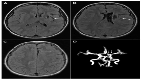
| Figure 5: Axial MRI of the brain, 12 month post-stroke [A: Areas of cystic gliosis seen within the left parasagittal frontal lobe, left extreme capsule, left head of caudate nucleus, and within the right parasagittal frontal lobe region. The associated ex-vacuo dilatation of the left lateral ventricle and left sylvian fissure is also noted; B: Time-of-flight magnetic resonance angiography of the brain demonstrating stable narrowing of the left internal carotid artery and left middle cerebral artery (arrows)]. |