|
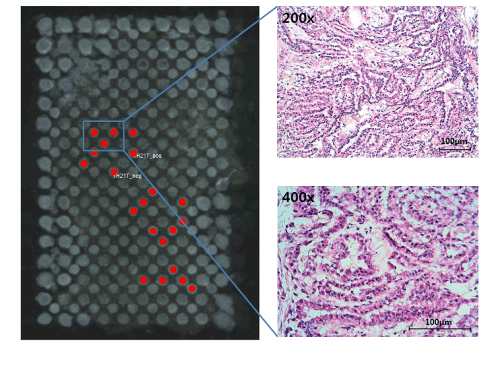
| Figure 1: General procedure. Representative optical image of the cryosection ITO slide with matrix (left) and magnified areas of the H&Estained consecutive cryosection slide of a tumor sample (right) are shown. The H&E-stained cryosection slide was evaluated for tumor-rich (>75%) area. Guided by the H&E-stained cryosection slide, deposited matrix spots on ITO cryosection slides representing tumor-rich area (shown in red) were selected using FlexImaging software for each tumor sample. Similar procedures were performed for normal tissue samples. Mass spectra data from selected spots was then exported to ClinProTools for further data processing. |