[A, C, E: xObj, 5, B, D: Obj.x20, F: Obj.x10]
 [A, C, E: xObj, 5, B, D: Obj.x20, F: Obj.x10] |
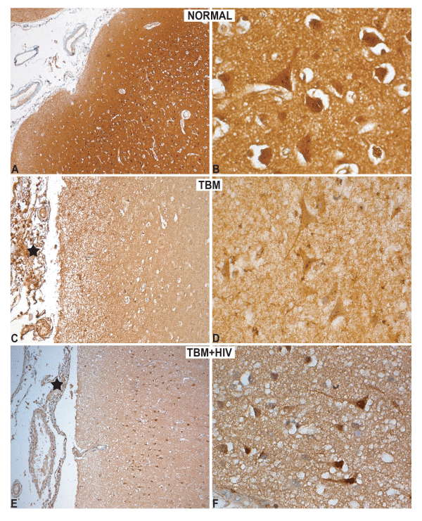
| Figure 8: Immunohistochemical labeling: HSPA8. A-B: Normal cortex shows intense labeling of neuropil of cortical ribbon (A) and neuronal soma (B). C-D: In cases of TBM, the labeling of both the neuropil (C) and neurons in grey matter is low (D) reflecting downregulation. The histiocytes in the exudates (C, asterix) are intensely labeled. E-F: In TBM with HIV, the neuropil labeling is very low (E) except for few large pyramidal neurons (F) that reveals nuclear labeling and pale dendritic arborization. Labeling is also seen of meningeal macrophages in the subarachnoid space (E, asterix). |