|
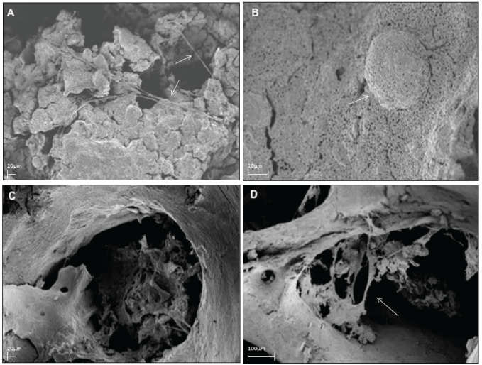
| Figure 3: A and B: SEM morphology analysis of BMMCs cultured on silicate granules. Cells appear to be well attached to the substrate with several pseudopodias and cytoplasmic extroflessions as indicated by arrows. A large amount of debris is present on the surface of cells grown on silicate granules. C and D: SEM morphology analysis of BMMCs cultured in presence of DMB. Cells form multi-aggregate localized into bone cavity with short pseudopodias. The arrows indicated pseudopodias. |