 |
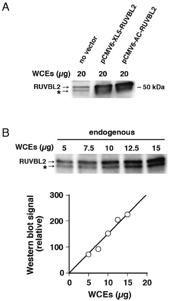
| Figure 1: Detection of endogenous RUVBL2. (A) Detection of RUVBL2 by Western blot analysis. WCEs of MRC5-SV cells without transfection and HEK293T cells transfected with a RUVBL2 expression vector (pCMV6-XL5- RUVBL2 or pCMV6-AC-RUVBL2) were separated by SDS-polyacrylamide gel electrophoresis. RUVBL2 protein on a blotted membrane was detected by anti-RUVBL2 antibodies. (B) Correlation between the amount of WCEs of MRC5-SV cells and the Western blot signal of endogenous RUVBL2. Upper panel: Western blot; lower panel: correlation between the amount of WCEs and the Western blot signal (average from two experiments). A cross-reacting protein in the lower band, which remains to be identified, is indicated by the asterisks in (A) and (B). |