|
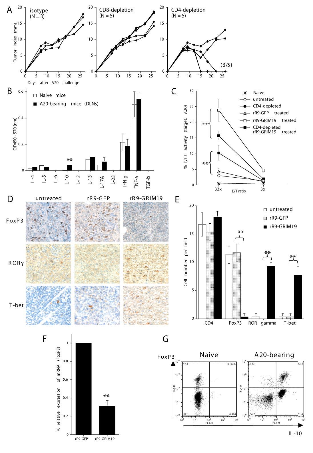
| Figure 2: IL-10-producing immunosuppressive CD4+ T cells were predominant in A20 tumor-bearing mice. (A): A20 tumor-bearing mice (n = 5 per group) were treated with i.p. injections of anti-CD4 (GK1.5) mAbs, anti-CD8 (53-6.7) mAbs, or control rat IgG (200 μg/mouse) on days 7 and 11. No other treatment was provided. Data are representative of three individual experiments. Data from only one representative experiment (tumor index from individual mice) is shown. (B): Cytokine-profile of CD4+ T cells from DLNs of A20-bearing mice on day 18 or control naïve mice with multiple cytokine ELISA analyses by stimulation with PMA plus ionomycin. Data are representative of two individual experiments (** p < 0.01). (C): CTL assay against A20 cells. A20 cells were subcutaneously injected into naïve mice on day 0. rR9-GRIM19 or rR9-control proteins were injected i.t. daily on days 9, 10, 11. Some A20 tumor-bearing mice were also treated with i.p. injections of anti-CD4 (GK1.5) mAbs on days 7 and 11. On day 28, DLN cells were collected and restimulated in vitro. On day 33, CTL reactivity with A20 cells was determined. The E/T ratio and the percent lysis activity are shown on the x and y axes, respectively. Data are representative of 2 individual experiments (** p < 0.01). (D-E): FoxP3+ cells were predominantly infiltrated in A20 tumor-bearing skin area, but were decreased after treatment of rR9-GRIM19. (D): A20-bearing mice were treated with i.t. injections of rR9-GFP or rR9-GRIM19. On day 13, skin biopsies were performed from the area including the A20-tumor mass. Vertical skin sections were stained with anti-CD4 mAb, anti-FoxP3 mAb, anti-RORγ Ab, or anti-T-bet mAb (original magnification x200). Data are representative of two individual experiments. (E): A summary of the number of cells infiltrating A20-tumor areas is shown in Fig. 2D. The average number of positive cells, based on immunohistochemical staining with the indicated mAbs, was counted per cell-infiltrated field (400) at three separate areas from each skin section. (**; p < 0.01). (F): Expression of FoxP3 mRNA in skin samples also decreased after intratumoral injections of rR9-GRIM19. Real-time PCR was performed using homogenized skin samples from A20 tumor area on day 13. Relative expression of FoxP3 mRNA to GAPDH mRNA was measured. Data are representative of two individual experiments. Data from only one representative experiment is shown. (G): IL-10-producing CD4+ T cells in DLNs from A20 tumor-bearing mice were observed in both FoxP3+ and FoxP3- cells. The CD4+ T cells from DLNs of untreated A20 tumor-bearing mice (on day 20) or from naïve mice were restimulated in vitro with PMA and ionomycin, and intracellular staining was performed with mAbs. Data are representative of three individual experiments. Data from only one representative experiment is shown. |