 |
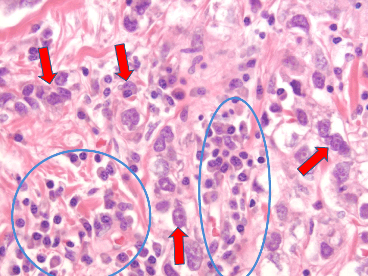
| Figure 1: This figure demonstrates the histology of a breast ductal adenocarcinoma (H&E, X400). Malignant breast cells are indicated by the red arrows. Intermixed inflammatory cells are indicated by the blue circles. Of note, malignant cells cannot be separated from inflammatory cells by macrodissection because there is no clear line of separation. |