|
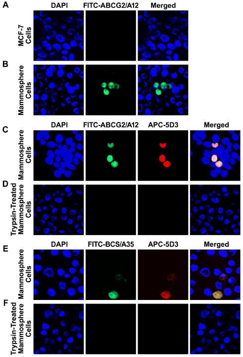
| Figure 3: The cells stained with ABCG2-specific aptamer ABCG2/A12 or ABCG2-specific mAb can form mammospheres. A. ABCG2/A12 did not recognize the differentiated breast cancer MCF-7 cells: left section, cells were stained with DAPI; middle section, with FITC-labeled ABCG2/A12; right section, merge of the previous two sections. B. ABCG2/A12 recognizes the ABCG2-expressing BCS cells in mammospheres derived from MCF-7. C. ABCG2/A12 and ABCG2-specific mAb 5D3 recognized the same BCS cells in mammospheres: left section, cells were stained with DAPI; second section, with FITC-labeled ABCG2/A12; third section, with APC-conjugated ABCG2-specific mAb 5D3; right section, merge of the previous three sections. D. Short time trypsin digestion completely abolished the binding of ABCG2/A12 and ABCG2-specific mAb 5D3 to BCS cells in mammospheres. E. BCS cells in mammospheres derived from MCF-7 were co-stained with BCS/A35 and ABCG2-specific mAb 5D3: left section, cells were stained with DAPI; second section, with FITC-labeled BCS/A35; third section, with APC-conjugated human ABCG2-specific mAb 5D3; right section, merge of the previous three sections. F. Short time trypsin digestion completely abolished the binding of BCS/A35 and ABCG2-specific mAb 5D3 to BCS cells in mammospheres. G. FITC-ABCG2/A12 negative (#12-) cells (4,000), sorted out by FACS from mammospheres derived from MCF-7, can barely form mammospheres. H. FITC-ABCG2/A12 positive (#12+) cells (4,000), sorted out by FACS from mammospheres derived from MCF-7, can form mammospheres. I. APC-conjugated ABCG2- specific mAb 5D3 negative (5D3-) cells (4,000) can barely form mammospheres. J. APC-conjugated ABCG2-specific mAb 5D3 positive (5D3+) cells (4,000) can form mammospheres. K. Numbers of mammospheres formed from above four cell populations. BCS cells have significantly higher rate of clathrin-independent endocytosis than the differentiated breast cancer cells. A. Staining of the differentiated breast cancer MCF-7 cells with hTF in the absence of CDE inhibitor: top left, cells were stained with DAPI; bottom left, with Alexa fluor 633-conjugated hTF; top right, amplified image of the cells from bottom left section; bottom right, merge of the top left section and bottom left section. B. Staining of the differentiated breast cancer MCF-7 cells with hTF in the presence of CDE inhibitor MDC. C. Staining of the differentiated breast cancer MCF-7 cells with hTF in the presence of CDE inhibitor sucrose. D. Staining of the mammosphere cells derived from MCF-7 with BCS/A35 in the absence of CDE inhibitor: top left, cells were stained with DAPI; top right, with FITC-labeled BCS/A35; bottom left, merge of the top left section and top right section; bottom right, amplified image of the cells from bottom left section. E. Staining of the cells derived from mammospheres with BCS/A35 in the presence of CDE inhibitor MDC. F. Staining of the cells derived from mammospheres with BCS/A35 in the presence of CDE inhibitor sucrose. G. Co-staining of the cells derived from mammospheres with BCS/A35 and hTF in the absence of CDE inhibitor: top left, cells were stained with DAPI; top right, with FITC-labeled BCS/A35; bottom left, with Alexa fluor 633-conjugated hTF; bottom right, merge of the previous three sections. H. Co-staining of the cells derived from mammospheres with BCS/A35 and hTF in the presence of CDE inhibitor MDC. I. Co-staining of the cells derived from mammospheres with BCS/A35 and hTF in the presence of CDE inhibitor sucrose. |