 |
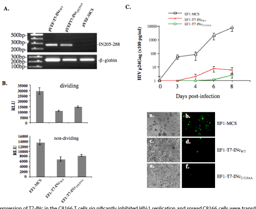
| Figure 4: Stable expression of T7-INc in the C8166 T cells significantly inhibited HIV-1 replication and spread. C8166 cells were transduced with lentiviral vector (pYEFP-MCS) or vector harboring T7-INc or T7-INc215, 9AA (pYEFP-T7-INc or T7-INc215, 9AA) and selected by puromycin (0.5μg/ml) for 7 days A: The presence of T7-INcwt or T7-INc215, 9AA mRNAs in C8166 stable cell lines was detected by RT-PCR using specific primers, as described in Materials and Methods (upper panel). Meanwhile, the human globin mRNA level from each RNA sample was monitored by RT-PCR (lower panel). B: The aphidicolin-treated (lower panel) or non-treated (upper panel) T7-INc expressing C8166 cell lines were infected with equal amounts of HIV-1 pNL4.3 Luc+/env+. At 48 h post infection, equal amounts of cells were collected and the luc activity was measured. C. pNL4.3-GFP virus at 0.01 MOI were used to infect vector-, T7-INcWT, or T7-INc215, 9AA transduced C8166 cell lines. At different time points, the supernatants were collected and HIV Gag-p24 levels were measured to monitor virus replication level (upper panel). Values were representative of two independent experiments. At day 5 post-infection, the HIV-infected (GFP-positive) cells were observed under fluorescence microscope (lower panel). |