 |
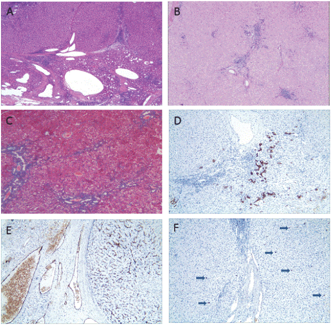
| Figure 3: (A-B) Well-defined, non-encapsulated tumor, composed of nodular hepatocytes with “cirrhosis-like” architecture, separated by fibrous septa. Numerous duct-like structure proliferations with scattered lymphocytes (hematoxylin & eosin, 40X), (C) Architectural disturbance caused by fibrous septa linking portal tracts (Masson staining, 100X), (D) Immunostaining for CK7 shows ductular reaction and phenotypic switching of the hepatocytic cytokeratins, reflecting long-standing intrahepatic cholestasis (CK7 immunostaining, 100X), (E) Immunostaining for CD34 shows a large sum of vascular endothelial cells which proliferated widely (CD34 immunostaining, 100X), (F) Immunostaining for Ki-67 shows the lower expression of proliferation (<1%, Ki-67 immunostaining, 100X). |