 |
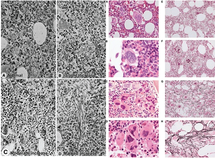
| Figure 1: Grading of reticulin fibrosis (RF, silver impregantion stain) according to Ellis et al. [11] in polycythemia vera (PV, left pannels black-white). Comparison of bone marrow histology and grading of RF according to Wilkins et al. [22]: A vs E: normocellular ET with small medium to large pleomorphic megakaryocytes and reticulin fibrosis grade 0/1, B vs F atypical large cell ‘artefact’ in B and RF grade 2 around clustered megakaryocytes with hyperlobulated nuclei. C vs G dense clustered large megakaryocytes with hyperlobulated nuclei and RF grade 3. D vs H dense clustered immature megakaryocytes with large immature (dysmorphic) cloud-like hypolobulated nuclei and RF grade 4 [22]. |