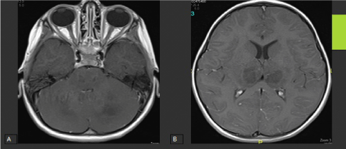
Figure 6:
Axial contrast-enhanced T1W1 MRI of the brain at the level of the pons (A), Basal Ganglia and Thalami (B) showing no evident post-contrast enhancement.