 |
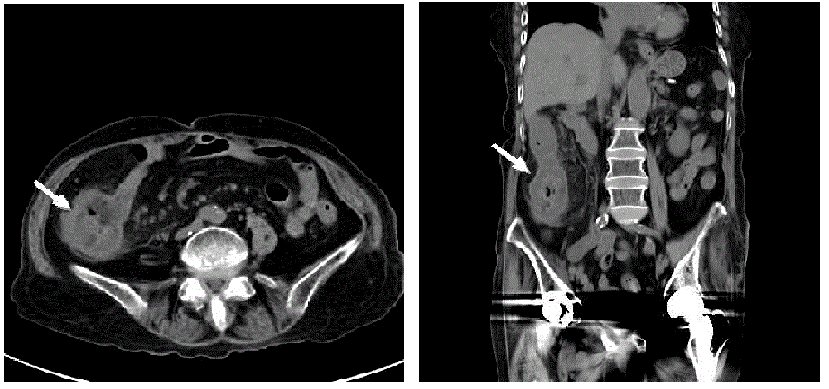
| Figure 1: Abdominal non-enhanced CT images (A) Transverse section at the level of ileocecal junction shows circumferential wall thickening of cecum with submucosal edematous change and pericolic infiltration (arrow). (B) Coronal section shows circumferential bowel wall thickening from cecum to ascending colon (arrow). |