 |
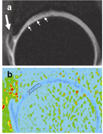
| Figure 2: An example case with cartilage damage: (a) A sagittal T1 weighted image (TR/TE = 700 ms/9 ms) with FATSAT. The anterior half of the acetabular cartilage is thinned and irregular (small arrows). Abnormal morphology of the anterior-superior labrum can be seen (large arrow). (b) The corresponding T1 map with an ROI in the cartilage. |