 |
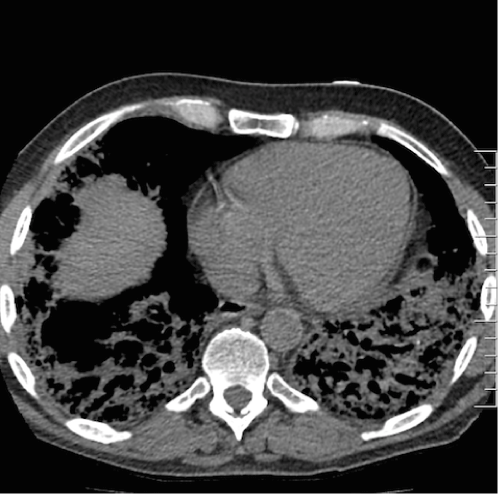
| Figure 2: CT chest with Contrast. Demonstrates in the right lung mild apical fibrosis, mild subpleural reticulation in the right upper lobe, moderate opacity in the right middle lobe with some traction bronchiectasis representing interstitial thickening. Severe opacity in the right lower lobe consisting of reticulation, traction bronchiectasis, likely some subpleural honeycombing. The left lung showed mild subpleural reticulation in the left upper lobe, moderately severe opacity in the lingula consisting of reticulation, and severe opacity in the left lower lobe similar to the right lower lobe. |