|
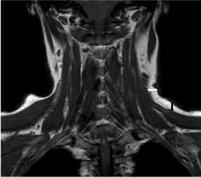
| Figure 3a: T1W MRI images of origin and insertion of the anomalous left neck muscle. Coronal image (a) showing its superior portion (white arrow) that fans out and merges with upper fibres of the left trapezius muscle (black arrow). Superomedially, it inserts into the left nuchal/occipital region (not shown). |