 |
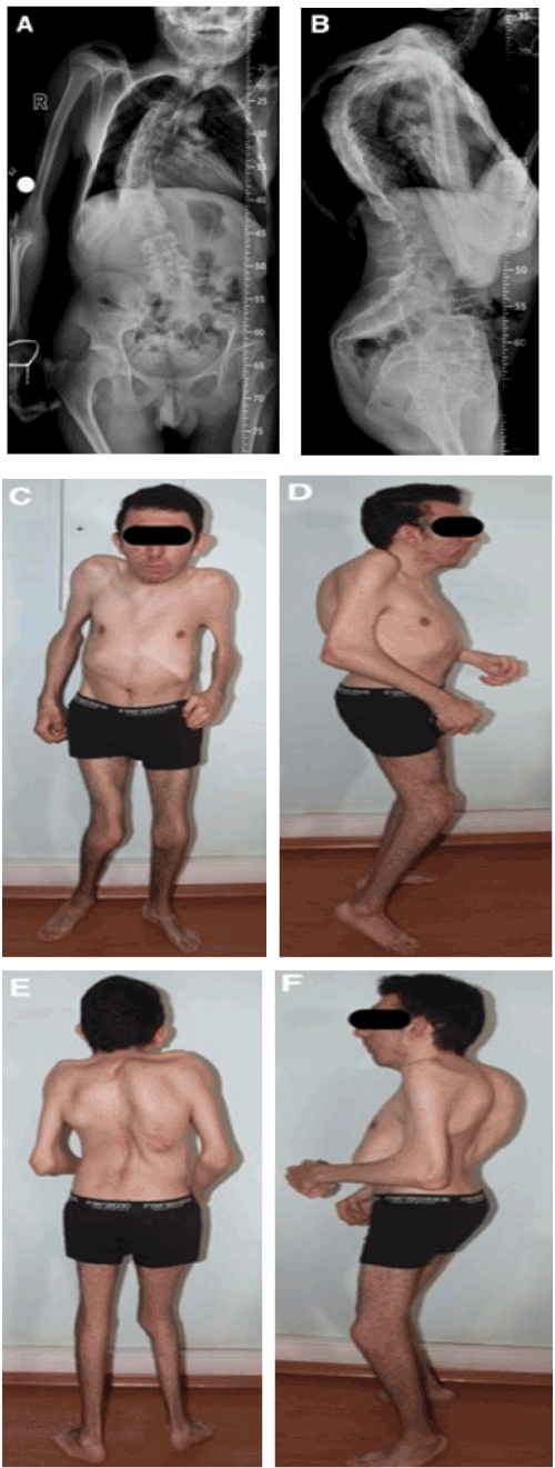
| Figure 1: The first case, a 17-year-old male patient whose brother also hadEscobar syndrome. Progressive scoliosis, kyphosis, ptosis, mild deafness,facial cranial dismorphism, low-set ears, low-set hairline, micrognathia, jointcontractures (axilla, elbow, hip, knee, ankle, neck); limitation of movement,hip dislocation, multiple pterygiums, vertical talus, camptodactlia,hypertrophy of gingiva, and tethered cord were identified. The surgicaltreatments of the patient included bilateral CVT and hip dislocation for openreduction, and inguinal hernia. Due to the progress of the kyphoscoliosisdeformity, he was treated with posterior spinal instrumentation and fusion.The instrumentation was removed due to problems caused by the implantin the third postoperative year. Radiographic image of the spinal deformityin the coronal (A) and sagittal plane (B). The appearance of the final examination, coronal and sagittal clinical pictures (C,D,E, and F). |