 |
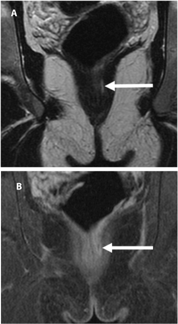
| Figure 4: Intersphincteric fistula in a 20-year-old man with Crohns disease. Coronal T2 image (A) demonstrates a linear focus of high signal intensity in the left intersphincteric region (arrow), with associated enhancement of the fistulous track on post-contrast image (B). |