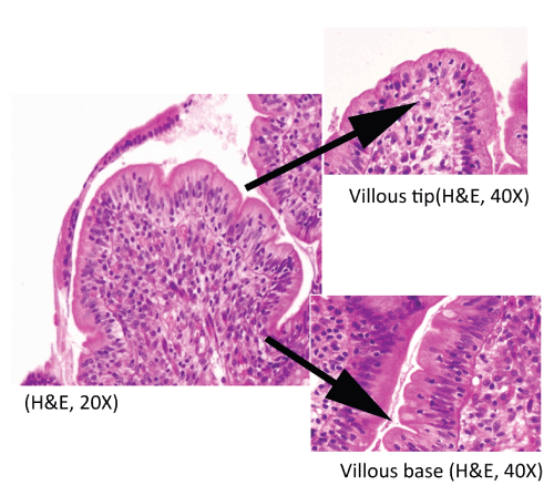
Figure 3:
Duodenal intraepithelial lymphocytosis (75/100 enterocytes) with loss of decrescendo pattern of intraepithelial lymphocyte distribution from villous tip to the base.