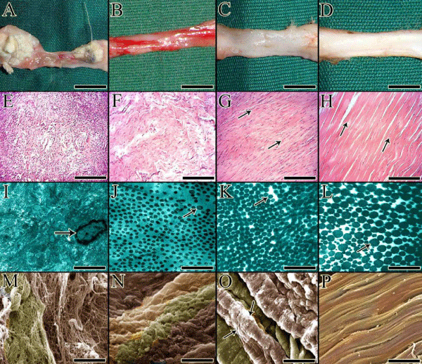
(A to D) are gross morphologic Figures, (E to H) are light microscopic micrographs, (I to L) are transmission electron ultramicrographs and (M to P) are scanning electron micrographs. (A, E, I and M) show an injured healing tendon in the inflammatory phase of tendon healing. (B, F, J and N) show an injured healing tendon in the fibroplasia phase of tendon healing. (C, G, K and O) show an injured healing tendon in the early remodeling phase of tendon healing. (D, H, I and P) show an injured healing tendon in the late remodeling phase of tendon healing. In the inflammatory phase of tendon healing the injured tendon is inflamed and the tendon edges are necrotized (A). The inflammatory cells consisting of neutrophils, lymphocytes and macrophages are infiltrated in the defect area and start to lyse the necrotic tissues (E). By transmission electron microscopy (I), a macrophage (arrow) could be seen in the injured area and the ultrastructure of the injured area is highly amorphous and no fibrillar pattern could be detected because most of the collagen fibrils of the scaffold have been lysed and no characteristic fibrillogenesis has still occurred. Under scanning electron microscopy the density of the collagen fibrils is much low and most of the tissue spaces between the collagen fibrils are free of collagen fibrils. In fibroplasia, the vascularity of the tissue is high and the injured area of the tendon is hyperemic (B), the collagenous matrix is formed but it is immature in nature because the collagen fibers are not still formed but the number of collagen fibrils is gradually increasing (F). Generally the tissue is hypercellular and the density of the collagenous mass is low (F). At ultrastructural level, the immature collagen fibrils have newly developed (arrow) but their density is low (J). Under scanning electron microscopy (N) the collagen fibrils have developed but they have not still developed to collagen fibers. At early remodeling phase, the hyperemia have reduced and the density of the new tissue has increased but still is not comparable to normal (C). The collagen fibers are recently formed and are unidirectionally aligned (G, arrows). At ultrastructural level, the density and diameter of the collagen fibrils have increased (K) so that the collagen fibers are formed which could be well detected under scanning electron microscopy (O, arrows). In the late remodeling phase, the density of the new tendon have greatly increased (D), and the bundles of collagen fibers are clearly detectable (H) and are highly aligned (H). Under transmission electron microscopy (I) the collagen fibrils are distributed in multimodal pattern so that more than three different categories of collagen fibrils could be detected based on their transverse diameter (I). Under scanning electron microscopy, the collagen fibers that have well differentiated and are densely packed could be seen. Scale bar: A to D: 1 cm, E to H: 50 μm, I to L: 900 nm, M to P: 2 μm.