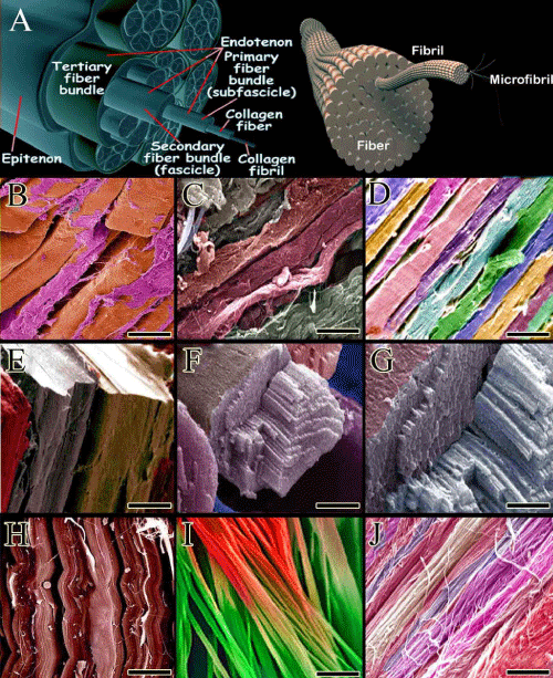
The hierarchical architectures of tendon and ligaments and their tridimensional architectures have significant similarities except that ligaments are denser than tendons and their collagen fibers are twisted. The collagen molecules are polymerized as tropocollagen, micro fibrils, fibrils, fiber bundles and fascicles (A). Endotenon covers the collagen fibers and bundles of collagen fibers internally and epitenon covers the tertiary fiber bundles (the largest bundles of collagen fibers). All these architectures are covered by paratenon in extra-synovial tendons and by tenosynovium or tendon sheath in intra-articular tendons (A). Ligaments (B) are denser than tendons (C, D). (E) is a scanning electron micrograph (SEM) of a normal tendon fascicle. (F) shows a large fiber bundle in which the collagen fibers are densely packed and formed a collagen fiber bundle (F and G). (H) is an ultramicrograph by SEM of the ligament fibers. Normally, these fibers are highly dense and aligned in a unidirectional pattern. (I and G) are the collagen fibrils. The collagen fibrils are aligned in one direction and aggregation of the highly aligned collagen fibrils produces the collagen fibers. B-D and H-J are longitudinal sections while E-G are sagital and cross sections. The ultramicrographs are colored through the structure detection software. Scale bar for B-D: 10 μm, E:100 μm, F:45 μm, G:22 μm, H: 10 μm, I: 700 nm, J: 1.8 μm.