|
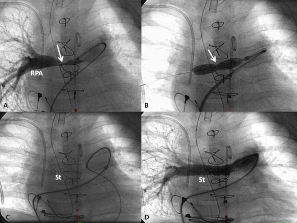
| Figure 3: Selected cineangiographic frames from pulmonary artery cineangiogram demonstrating severe narrowing (solid arrow in A) in the right pulmonary artery (RPA). During implantation of the stent (B) a waist of the balloon was seen (solid arrow in B) which was completely abolished following further inflation of the balloon. Following the removal of the balloon, the stent (St) is seen and is widely open (C). Follow-up angiogram shows wide open St in the RPA (D). Multiple radio-opaque structures such as catheters, sternal wires, pacemaker wires and endo-tracheal tube are seen and are not germane for the stent implantation result. |