 |
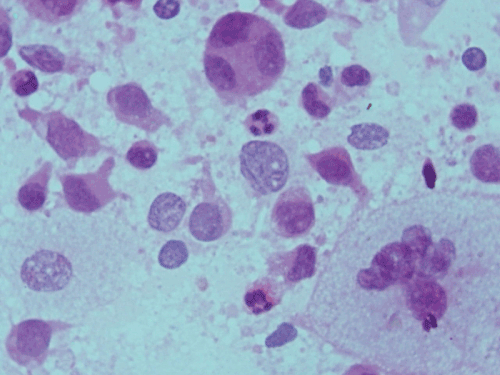
| Figure 3: Cytosmears show diagnostic Langerhans cells admixed with eosinophils, histiocytes, polymorphs and giant cells. Langerhans cells are characterized by oval to reniform nuclei and abundant pale cytoplasm. The nuclei show prominent grooving, finely granular chromatin pattern and inconspicuous nucleoli. (H&E x40). |