|
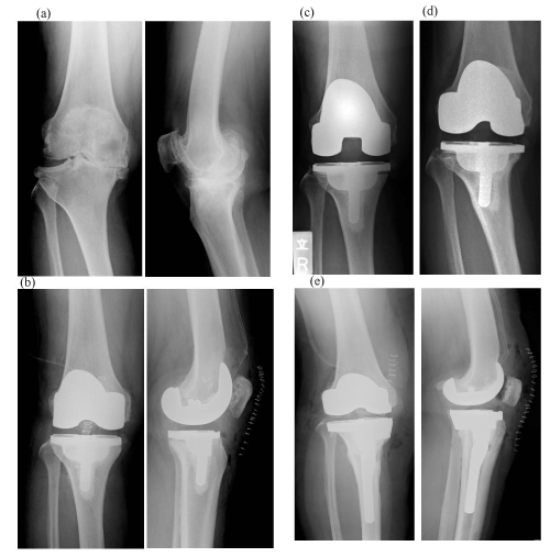
| Figure 1: Case 1. (a) Plain radiographs taken prior to primary TKA surgery. (b) Plain radiographs taken after the primary TKA surgery. Tibial component was implanted with 4 degrees of varus and 4 degrees of posterior slope.(c)Three months after primary TKA. Radiolucency below the tibial component was clearly observed. (d) Eight months after primary TKA. A sinking of the tibial component and varus deformity had occurred. (e) Approximately 2.5 years after initial TKA, tibial component revision was performed. |