 |
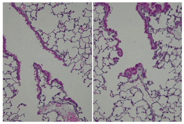
| Figure 5: Hematoxylin and eosin stain for the alteration of the epithelial cells of terminal bronchioles. (Left) The filtered air group. Note the centriacinar regions of alveolar ducts were generally straight and thin alveolar walls. (Right) The HONO exposure group. Note the hyperplasia of the terminal bronchial epithelial cells with the meandering irregularly and without dysplasia. |