 |
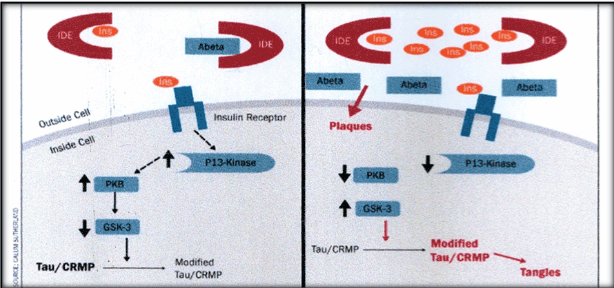
| Figure 1: Figure to left represents insulin signaling under normal healthy conditions: insulin adapts in receptor, activates PI3-K/Akt(PKB) and inhibits GSK3 in the intracellular compartment, with good balance between insulin, IDE and Aβ in extracellular space. Figure to right, represents states of insulin resistance and AD pathology: insulin remains in extracellular space competing for IDE favoring deposition of Aβ. In the intracellular compartment, PI3K fails to activate and promotes GSK3 activity with tau phosphorylation and deposition of tangles. |