 |
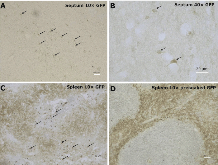
| Figure 1: Photomicrographs of IHC stained GFP-positive cells in the septum (A and B; most GFP+ cells are in the lateral septum, with fewer in the medial septum), and the spleen (C) in IL2p8-GFP mice. Panel D illustrates that the reactivity of primary antibody was quenched in the spleen with 5 μg/ml free recombinant GFP protein prior to use in staining protocol. |