 |
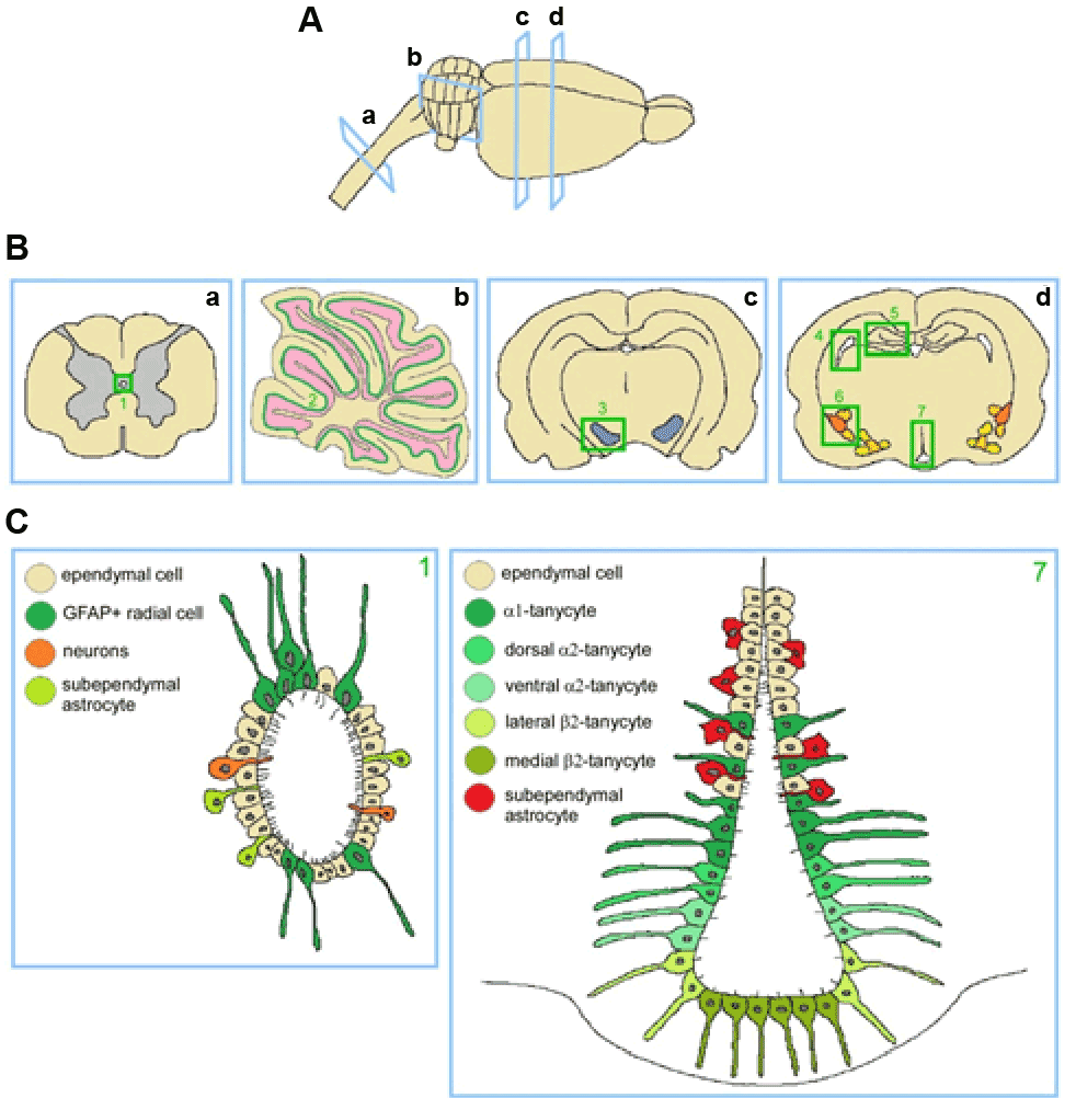
| Figure 1: Classical and unconventional neurogenic niches. A: longitudinal view of a rat brain and part of the spinal cord, indicating cross sections. B: Cross sections of spinal cord (a), cerebellum (b) and rat brain (c, d) at different levels. Regions where neurogenic potential or NSC presence has been demonstrated are highlighted in green. Spinal cord central canal (1), Purkinje cell layer (2), substantia nigra (3), SVZ of the lateral ventricles (4), SGZ of the dentate gyrus (5), amygdala (6) and hypothalamus (7). C: Cellular composition of the newly described neurogenic niches: central canal (left panel) and hypothalamus (right panel). |