 |
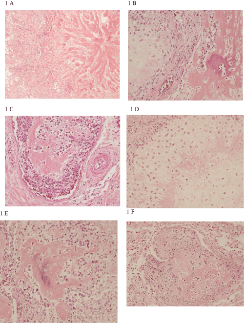
| Figure 1: Primary mammary combined osteosarcoma with low-grade central parts (to the right), showing a large amount of bone matrix and a high-grade more cellular periphery (to the left), (A), (objective x 4). Another part of the primary tumour demonstrated presence of cartilage to the left and bone to the right (B), (objective x 20). Tumour invasion with bone matrix formation even within the intravascular metastasis was seen (C), (objective x 20). Cartilage (D) and bone (E) also appeared in the lung metastases (objectives x 20). Abundance of bone matrix was also seen in the lung vessel metastases (F), (objective x 20). Light micrographs of dog No. O389 with haematoxylin and eosin (HE) staining. |