 |
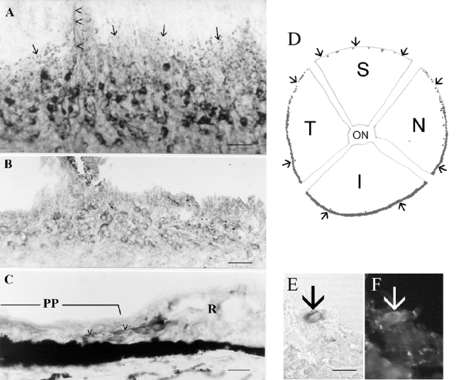
| Figure 1: A. Retinal whole mount (NADPH-d) showing numerous stained cells next to the oraserrata (marked by arrows). At one place, stained processes run into the pars plana region (arrowheads). B. Tangential section stained with antibodies against nNOS at the same position. C. NADPH-d. Sagittal section showing stained processes (arrowheads) of the positive neurons towards the pars plana (PP) region. R=retina. D. Schematic drawing of a whole mount showing the different distribution of NADPH-d/nNOS positive cells (black dots) in the porcine retina. T=temporal, S=superior, N=nasal, I=inferior quadrant. ON=optic nerve region. Arrows mark the oraserrata. E, F. Double-staining of a single cell (arrow) with NADPH-d (E) and Calretinin (F). Scale bar in A and B: 50 μm, in C, E and F: 20 μm. |