|
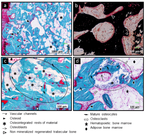
| Figure 8: Histological images of defects implanted with biomaterial showing an advanced stage of bone regeneration. (a) Well-mineralized regenerated trabecular bone (blue) with osteogenic activity and vascular channel systems surrounded by osteoid lines (red). (b) Cubic active osteoblasts associated to wide osteoid lines at well mineralized trabecular bone surface. (c,d) Circular areas showing osteointegrated remains of partially resorbed granulates surrounded by mineralized bone with vascular channels and osteocytes. (d) Remodelation of new bone by osteoclast and resorption of osteointegrated granulates by osteoclast at their bone-free surface in contact with bone marrow. (a,c,d) Stained with Goldner’s trichrome technique showing mineralized bone in blue and collagen fibers present in non-mineralized tissue in red, corresponding to connective tissue outside defect and to non-mineralized organic bone matrix (osteoid) inside defect. (b) Stained with Von Kossa showing mineralized bone in black and non-mineralized organic bone matrix (osteoid) in red. |