 |
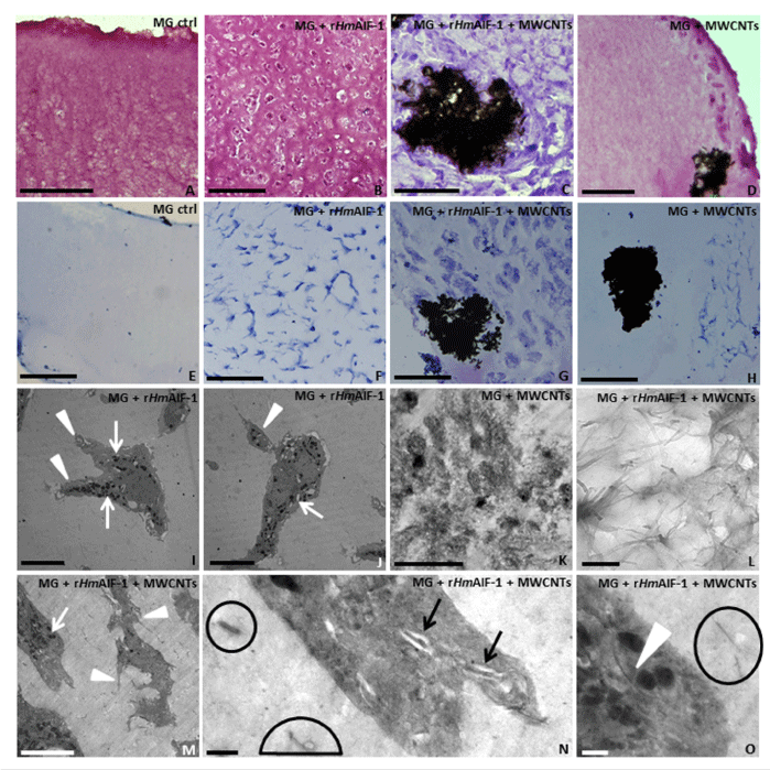
| Figure 1: Morphological and colorimetric analyses of cells infiltrating the MG removed from the animal 1 week after injection. (A-H) Appearance under the light microscope of the cells infiltrating the gels without (A, E) and with the rHm AIF-1 (B, F). The rHmAIF-1 recruits a large number of cells (B) positively stained by May GrunwaldGiemsa method (E, F). (C, D, G, H) in MG pellet supplemented with both MWCNTs and rHmAIF-1 (C, G) or only with MWCNTs (D, H), migrating cells positive for GrunwaldGiemsa staining (G, H) are visible dispers ed in the biomatrix and surrounding the MWCNTs aggregates (arro wheads). (I-O) TEM images. (I, J, M-O) Detail of cells infiltrating the MG pellets characterized by a cytoplasm with numerous highly electron-dense granules (arrows), pseudopodia (arrowheads in I, J) and lamellipodia (arrowheads in M). (K, L, N, O) Detail of MWCNTs dispersed or grouped in aggregates differently sized inside the MG sponge (K, L, encircled in N, O) or in intracellular vesicles (arrows in N) and freely dispersed in the cytoplasm (arrowhead in O). Bars in A-H: 50 μm; Bars in I-K, M: 5 μm; Bars in L: 200 nm; Bars in N, O: 500 nm. |