 |
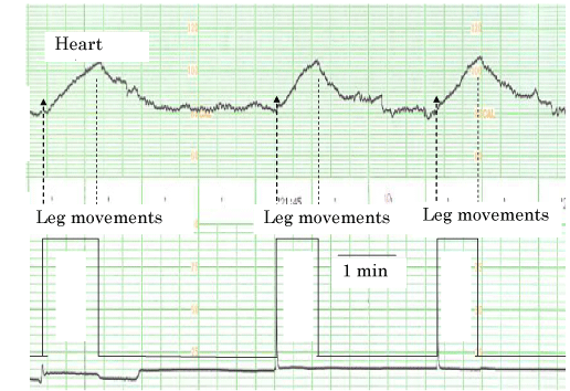
| Figure 8: FHR acceleration was simulated in adult. Heart rate was recorded placing the heart probe on anterior chest wall and the legs were moved continuously on the chair for 1 minute, and heart rate was recorded by an ultrasonic fetal monitor. The onset and stop of movements were marked on the chart by touching the sensor of uterine contraction. Triangular heart rate changes similar to fetal accelerations were recorded, but the heart rate change was not recognized by the subject person. Therefore, the center for the LTV and acceleration will be located not at the brain cortex, but maybe in the midbrain. |