|
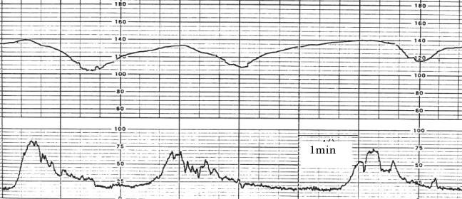
| Figure 3: Intrapartum FHR (upper) and uterine contraction (lower) of a late deceleration with the loss of LTV. ‘FHR tracing is very flat comparing to the 3 FHR tracings listed in figure 1. The LTV amplitude in the figure was less than 1 bpm, and the frequency power spectrum data are lower than that of FHR in fetal resting state [7]. FHR acceleration is also lost. Neonatal Apgar score was 3, showing severe neonatal asphyxia. |