 |
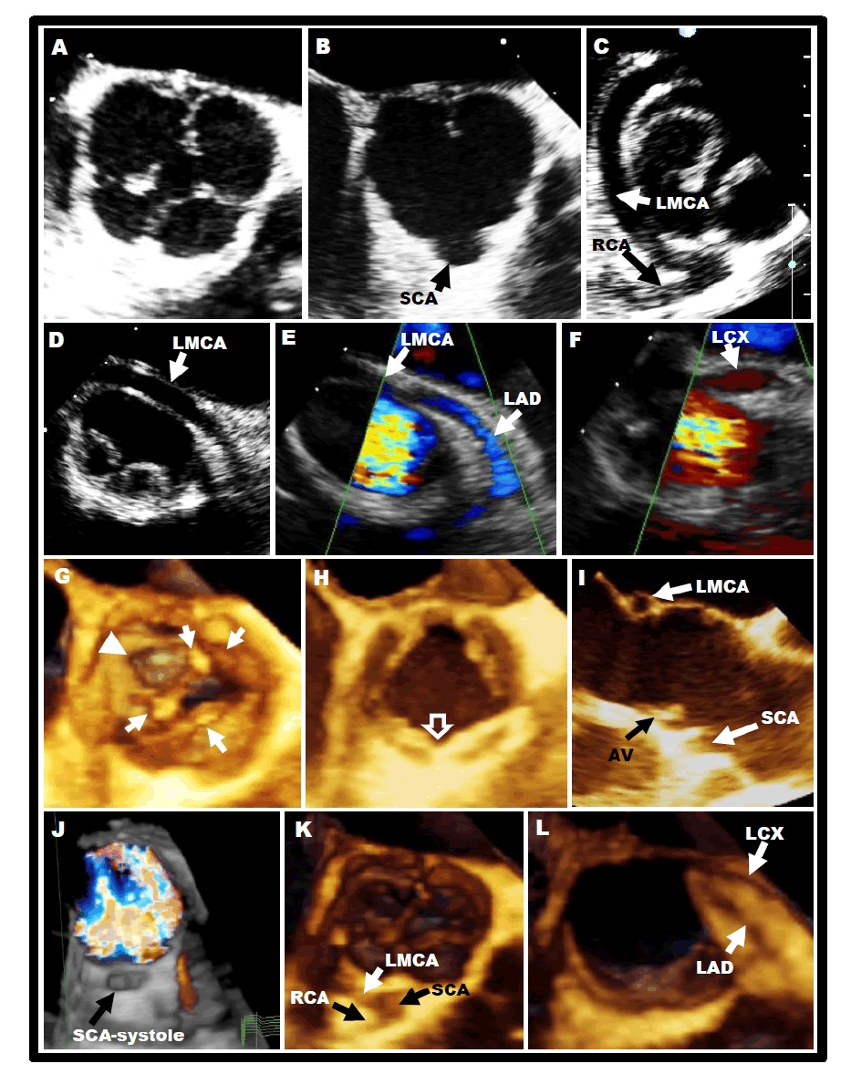
| Figure 1: Echocardiographic findings. 2D-TEE. (A) Short axis of the QAV. (B) The SCA is seen originating from the “right coronary sinus” giving rise to the RCA and LMCA (C) before dividing into LAD and LCX arteries (E and F respectively). 3D-TEE. (G) corpus arantii (arrows) on the 4 valve leaflets, a leaflet fenestration (arrow head) and (H) short raphe (open arrow) are noted. (I) 3D reformat image demonstrating the high takeoff of the SCA (just above the sinutubular junction). A cross section of the LMCA is also noted. (J) 3D color image, showing the ostium of the SCA during systole. (K) Branching of the LMCA into LAD and LCX (L). 2D-2-dimensional; 3D-3 dimensional; SCA- single coronary artery; TEE-transesophageal echocardiography; LAD-left anterior descending; LCX-circumflex artery; LMCA-left main coronary artery; QAV-quadricuspid aortic valve; RCA-right coronary artery. |