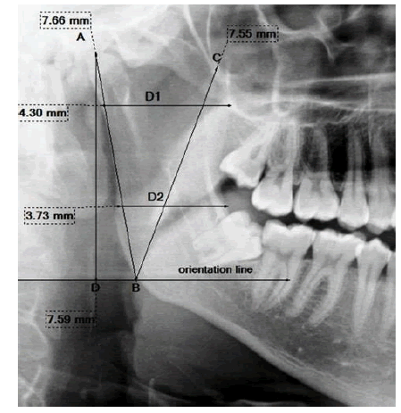
Figure 1:
The five linear ramus measurements performed on the digital panoramic image (D1: upper ramus breadth, D2: lower ramus breadth, AB: condylar ramus height, BC: coronoid ramus height, AD: projective ramus height).