 |
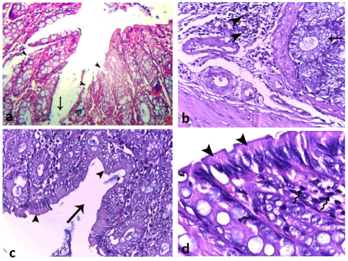
| Figure 3: Photomicrographs of NAC treated group (a,b) and ginger treated group (c,d) showing: (a) dilated intestinal crypts (arrows) and areas of sloughed and interrupted epithelium (arrow heads) among normal surface epithelium. (b) Dilated intestinal crypts lined by vacuolated cytoplasmic cells (arrow) and submucosal inflammatory infiltration (arrow head). (c,d) Intact intestinal epithelium (arrow heads) and dilated crypts (arrow). There are few inflammatory cells (wavy arrows). (H & E, (a,b,c) 400X; (d) 1000X). |