|
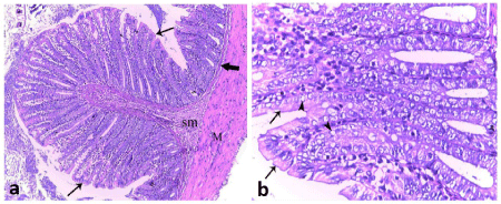
| Figure 1: Photomicrographs of sections in the colon of control group showing: (a) the mucosa arranged into deep narrow spaced intestinal crypts, lined by simple columnar epithelial cells (arrows), resting on a thin continuous layer of muscularis mucosa (thick arrow). Narrow submucosal layer (sm) separates the mucosa from the muscle layer (M) and extends in the core of the colonic mucosal folds. (b) The intestinal crypts are lined with both simple columnar epithelium (arrow) and Goblet cells (arrow heads). (H & E, (a) 100X; (b) 400X). |